File:The effect of embryo manipulation on 5meC staining in zygotes z3332337.jpeg
Original file (662 × 812 pixels, file size: 267 KB, MIME type: image/jpeg)
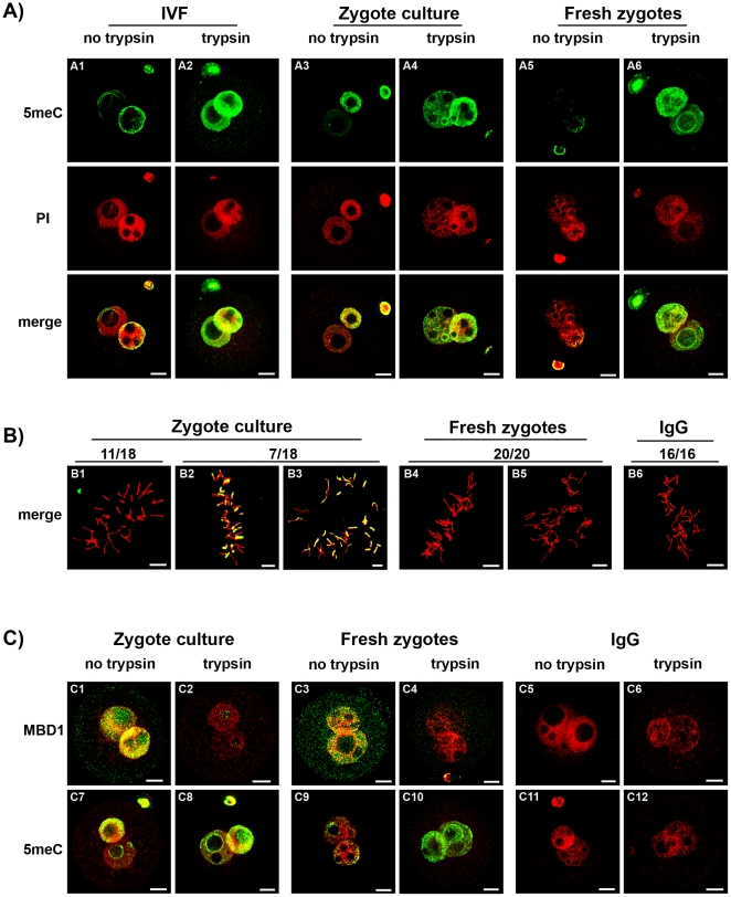
Asymmetric anti-5meC staining of the male and female pronucleus after acid-pretreatment has been reported [5], [6], [20], [21] yet was not confirmed by this study. The zygotes used in past studies were commonly generated by in vitro fertilization or subjected to culture in vitro (which provides logistic advantages for the feasibility of such studies). After antigenic unmasking with acid, the smaller (female) pronucleus in zygotes produced by in vitro fertilization (Fig 7A1) and or cultured in vitro (Fig 7A3) showed more anti-5meC staining compared to those collected directly from the oviduct (Fig 7A5). After antigenic unmasking by acid and trypsin, however, high levels of anti-5meC staining were consistently observed in both pronuclei of IVF (Fig 7A2), cultured (Fig 7A4) and fresh PN5 zygotes (Fig 7A6). Analysis of metaphase zygotes showed that culture from the early zygote stage caused variable levels of anti-5meC staining to persist in acid-only treated zygotes (Fig 7B). The level of methylation was assessed further by comparing staining with anti-MBD1 and anti-5meC in fresh and cultured zygotes (Fig 7C). This analysis showed that a similarly high level of MBD1 staining was observed in PN5 cultured (Fig 7C1) and fresh (Fig 7C3) zygotes, yet 5meC staining persisted in an asymmetrical fashion in cultured (Fig 7C7) but not fresh (Fig 7C9) zygotes. After acid and trypsin unmasking the MBD1 staining was lost from both treatments (Fig 7C2,4) and resulted in a similarly high level of staining with anti-5meC in both cultured and fresh zygotes (Fig 7C8,10). No staining was detected with non-immune control antisera for either antibody (Fig 7C5,6 and C11,12). The current results show that manipulation of the early embryo interferes with the maturational changes in zygotic chromatin that results in acid-resistant antigenic masking of 5meC, and this reduced level of masking was greatest in the female pronucleus giving an artifactual appearance of asymmetric demethylation.
Reference:
Li, Y., & O'Neill, C. (2012). Persistence of cytosine methylation of DNA following fertilisation in the mouse. PLoS One., 7(1), e30687. Epub 32012 Jan 30626.
Copyright Li, O'Neill. This is an open-access article distributed under the terms of the Creative Commons Attribution License, which permits unrestricted use, distribution, and reproduction in any medium, provided the original author and source are credited.
File history
Click on a date/time to view the file as it appeared at that time.
| Date/Time | Thumbnail | Dimensions | User | Comment | |
|---|---|---|---|---|---|
| current | 19:08, 7 August 2012 | | 662 × 812 (267 KB) | Z3332337 (talk | contribs) | Asymmetric anti-5meC staining of the male and female pronucleus after acid-pretreatment has been reported [5], [6], [20], [21] yet was not confirmed by this study. The zygotes used in past studies were commonly generated by in vitro fertilization or subje |
You cannot overwrite this file.
File usage
The following page uses this file: