File:The cell behaviours and its different locations down the AP embryonic axis.jpg
The_cell_behaviours_and_its_different_locations_down_the_AP_embryonic_axis.jpg (713 × 494 pixels, file size: 115 KB, MIME type: image/jpeg)
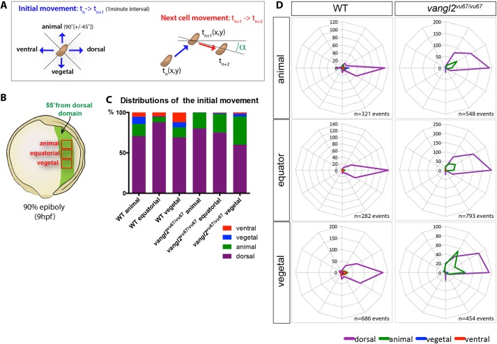
The cell behaviours and its different locations down the AP embryonic axis[1]
Image Title
There are 4 parts to this image - A, B, C and D. - Part A shows the initial movement of the cell and the next cell movement at 1 minute intervals. - Part B shows a late gastrula embryo, exploring the animal, equatorial and vegetal positions. - Part C is a graph of the distributions of the initial movement in WT and vangl2 embryos at 1 minute intervals at dorsal, animal, vegetal and ventral positions. - Part D shows the relative position of the next cell movement.
Image Copyright
Articles published in these journals are in the public domain and may be used and reproduced without special permission. However, anyone using the material is requested to properly cite and acknowledge the source.
- Note - This image was originally uploaded as part of an undergraduate science student project and may contain inaccuracies in either description or acknowledgements. Students have been advised in writing concerning the reuse of content and may accidentally have misunderstood the original terms of use. If image reuse on this non-commercial educational site infringes your existing copyright, please contact the site editor for immediate removal.
File history
Click on a date/time to view the file as it appeared at that time.
| Date/Time | Thumbnail | Dimensions | User | Comment | |
|---|---|---|---|---|---|
| current | 14:01, 18 August 2016 |  | 713 × 494 (115 KB) | Z5019526 (talk | contribs) | PMCID: PMC4510859 |
You cannot overwrite this file.
File usage
The following page uses this file:
