File:Thalassemia pic.jpg
Thalassemia_pic.jpg (440 × 367 pixels, file size: 31 KB, MIME type: image/jpeg)
Cells at cortical plate
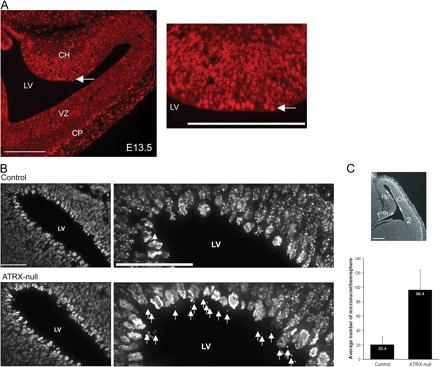
ATRX association with mitotic chromosomes and evidence of mitotic defects in ATRX-deficient neuroprogenitors in vivo. (A) ATRX staining of the cortex at E13.5. ATRX is highly expressed in all cells but is highest in the cortical hem (CH), which gives rise to hippocampal structures. ATRX is also expressed at increased levels in the differentiated neurons of developing cortical plate (CP). Mitotic cells that line the lateral ventricle are highly enriched for ATRX protein (arrows). Higher magnification of the cortical hem (right) demonstrates ATRX staining of mitotic chromosomes in cells that line the lateral ventricle (arrow). Punctate nuclear staining of ATRX in cycling cells of the ventricular zone (VZ) is characteristic of ATRX localization at PCH. (B) Cryosections obtained from control and ATRX null telencephalon were stained with DAPI to visualize mitotic chromosomes lining the lateral ventricle (LV) at E13.5. An increased incidence of micronuclei or dispersed chromosomes was detected in the vicinity of the mitotic layer (arrows) in the ATRX null embryonic brain. (C) The mean number of micronuclei or dispersed chromosomes were scored in the mitotic layer from point 1 to 2 as indicated by the white arrows (top) and are represented in the graph below (n = 4 for each control and ATRX null brain; P < 0.0001 by nonpaired t test). Error bars represent the standard deviation of counts from a total of 24 cortical hemisphere from a total of four brains. Cn, cortical neuroepithelium; Hh, hippocampal hem; Hp, hippocampal primordium. Bars: (A) 100 μm; (B) 40 μm; (C) 200 μm.
Loss of ATRX leads to chromosome cohesion and congression defects Kieran Ritchie, Claudia Seah, Jana Moulin, Christian Isaac, Frederick Dick, and Nathalie G. Bérubé Published January 28, 2008 // JCB vol. 180 no. 2 315-324 The Rockefeller University Press, doi: 10.1083/jcb.200706083 © 2008 Rockefeller University Press
File history
Click on a date/time to view the file as it appeared at that time.
| Date/Time | Thumbnail | Dimensions | User | Comment | |
|---|---|---|---|---|---|
| current | 11:02, 18 August 2011 |  | 440 × 367 (31 KB) | Z3217043 (talk | contribs) | Cells at cortical plate ATRX association with mitotic chromosomes and evidence of mitotic defects in ATRX-deficient neuroprogenitors in vivo. (A) ATRX staining of the cortex at E13.5. ATRX is highly expressed in all cells but is highest in the cortical h |
You cannot overwrite this file.
File usage
The following 2 pages use this file:
