File:Syngnathidae development 03.jpg
Syngnathidae_development_03.jpg (800 × 320 pixels, file size: 67 KB, MIME type: image/jpeg)
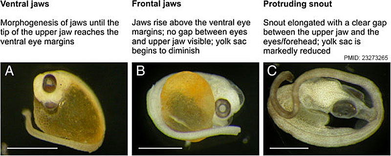
Figure 3 Snout formation
Descriptions of the three stages of the snout-formation period, along with examples for each stage. (A) S. abaster embryo (dechorionated) with ventrally developing jaws. (B) H. abdominalis larva with jaws rising frontally. (C) S. abaster larva with an elongated snout. Scale bars are 1 mm.
Reference
<pubmed>23273265</pubmed>| BMC Dev Biol.
Copyright
© Sommer et al.; licensee BioMed Central Ltd. 2012 This article is published under license to BioMed Central Ltd. This is an Open Access article distributed under the terms of the Creative Commons Attribution License (http://creativecommons.org/licenses/by/2.0), which permits unrestricted use, distribution, and reproduction in any medium, provided the original work is properly cited.
File history
Click on a date/time to view the file as it appeared at that time.
| Date/Time | Thumbnail | Dimensions | User | Comment | |
|---|---|---|---|---|---|
| current | 18:07, 19 January 2016 | 800 × 320 (67 KB) | Z8600021 (talk | contribs) |
You cannot overwrite this file.
File usage
The following page uses this file: