File:Streeter1957 plate01.jpg
Original file (1,500 × 2,009 pixels, file size: 486 KB, MIME type: image/jpeg)
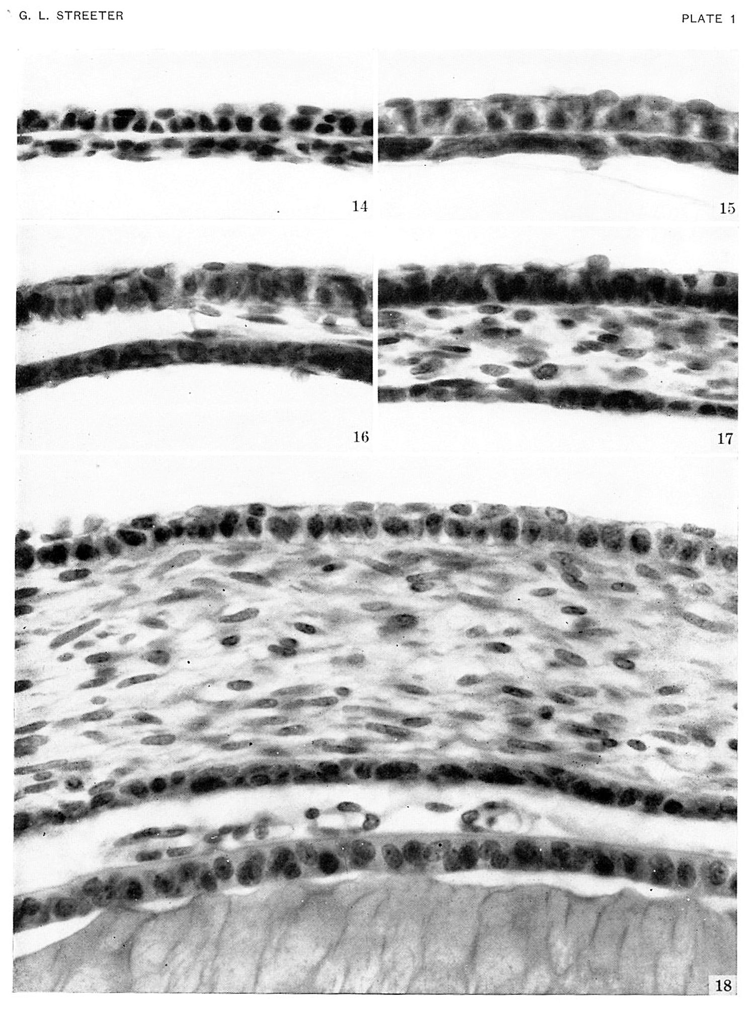
Plate 1. Photographs of the cornea in embryos stage 19-23
Photographs of the cornea in embryos belonging to horizons xix to xxiii.
A segment of the cornea near the mid-line is shown in an embryo of each horizon. The mesoclermal component is a thin layer one or two cells thick in the youngest stage. The stroma gmclually increases in thickness, and the cells at the inner border form a mesothelium. In the oldest specimen (Hg. 13), :1 part of the lens is included. The pupillary membrane is seen in the space between the mesothelium (Descement's) and the epithelium of the lens.
Fig. 14. No. 5609, xix, I5-I-I x 800.
Fig. 15. No. 431, xx, 37-2-2, x800.
Fig. 16. No. 1358F, xxi, 7-2-1, x800.
Fig. 17. No. 3681, xxii, 32-2-4, x800.
Fig. 18. No. 4570, xxiii, 70-3-4, x800.
| Historic Disclaimer - information about historic embryology pages |
|---|
 |
- Links: 1 Graph Embryos 11-23 | 4 Eye and optic nerve 19-23 | Plate 1 - Cornea | Plate 2 - Hypophysis
Reference
Streeter GL. Developmental Horizons In Human Embryos Description Or Age Groups XIX, XX, XXI, XXII, And XXIII, Being The Fifth Issue Of A Survey Of The Carnegie Collection. (1957) Carnegie Instn. Wash. Publ. 611, Contrib. Embryol., 36: 167-196.
Cite this page: Hill, M.A. (2024, April 24) Embryology Streeter1957 plate01.jpg. Retrieved from https://embryology.med.unsw.edu.au/embryology/index.php/File:Streeter1957_plate01.jpg
- © Dr Mark Hill 2024, UNSW Embryology ISBN: 978 0 7334 2609 4 - UNSW CRICOS Provider Code No. 00098G
File history
Click on a date/time to view the file as it appeared at that time.
| Date/Time | Thumbnail | Dimensions | User | Comment | |
|---|---|---|---|---|---|
| current | 16:29, 30 October 2016 | | 1,500 × 2,009 (486 KB) | Z8600021 (talk | contribs) | {{Streeter1957 figures}} Category:Week 8Category:Vision |
You cannot overwrite this file.
File usage
The following page uses this file: