File:Streeter1906 fig06.jpg

Original file (1,089 × 833 pixels, file size: 240 KB, MIME type: image/jpeg)
Fig. 6. Facial-acoustic Complex Human Embryo 7 mm
Sagittal section of a 7 mm. human embryo (B. 17), showing the relations of the facial-acoustic complex.
In Fig. 6 the ventral or motor division of the facial nerve can be seen cut obliquely at the ventral edge of the geniculate ganglion. In this embryo the dorsal root or pars intermedius is fully as large as the ventral or motor root; but the proportion is gradually reversed as one looks through older stages, due to the more rapid growth of the ventral root. The pars intermedius is in this sense a more prominent structure in fetal life than in the adult, indicating that phylogenetically it has played a more important role in lower forms than in man. With the development of the connective tissue the geniculate ganglion becomes inclosed in a sheath, and is walled off from the motor root, against which it continues to lie, as may be seen in Fig. 8.
| Historic Disclaimer - information about historic embryology pages |
|---|
 |
- Mall 1906 Links: Fig 1. 14mm Embryo | Fig 2. 30mm Embryo | Fig 3. Semicircular canal | Fig 4. Membranous Labyrinth | Fig 5. Acoustic nerve complex | Fig 6. Facial-acoustic Complex | Fig 7. Facial Nerve Pig Embryo 20 cm | Fig 8. Geniculate Ganglion | Plate 1. Human Embryo 4 to 20 mm | Plate 2. Human Embryo 30 mm | Membranous Labyrinth and Nerves
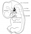
Fig 1 Membranous Labyrinth Human Embryo 14 mm
Fig 2 30mm Embryo
Fig 3 Semicircular canal
Fig 4 Membranous Labyrinth Growth
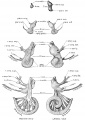
Fig 5 Acoustic nerve complex
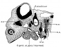
Fig 6 Facial-acoustic Complex Human Embryo 7 mm
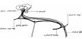
Fig 7 Facial Nerve Pig Embryo 20 cm
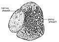
Fig 8 Geniculate Ganglion Human Embryo 30 mm
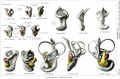
Plate 1. Membranous Labyrinth Human Embryo 4 to 20 mm
Plate 2. Membranous Labyrinth Human Embryo 30 mm


Reference
Streeter GL. On the development of the membranous labyrinth and the acoustic and facial nerves in the human embryo. (1906) Amer. J Anat. 6:139-165.
Cite this page: Hill, M.A. (2024, April 24) Embryology Streeter1906 fig06.jpg. Retrieved from https://embryology.med.unsw.edu.au/embryology/index.php/File:Streeter1906_fig06.jpg
- © Dr Mark Hill 2024, UNSW Embryology ISBN: 978 0 7334 2609 4 - UNSW CRICOS Provider Code No. 00098G
File history
Click on a date/time to view the file as it appeared at that time.
| Date/Time | Thumbnail | Dimensions | User | Comment | |
|---|---|---|---|---|---|
| current | 15:07, 31 July 2015 | | 1,089 × 833 (240 KB) | Z8600021 (talk | contribs) | |
| 14:59, 31 July 2015 |  | 1,316 × 947 (271 KB) | Z8600021 (talk | contribs) | ==Fig. 6. == {{Streeter1906 figures}} |
You cannot overwrite this file.
File usage
The following 12 pages use this file:
- Paper - On the development of the membranous labyrinth and the acoustic and facial nerves in the human embryo
- File:Streeter1906 fig01.jpg
- File:Streeter1906 fig02.jpg
- File:Streeter1906 fig03.jpg
- File:Streeter1906 fig04.jpg
- File:Streeter1906 fig05.jpg
- File:Streeter1906 fig06.jpg
- File:Streeter1906 fig07.jpg
- File:Streeter1906 fig08.jpg
- File:Streeter1906 plate01.jpg
- File:Streeter1906 plate02.jpg
- Template:Streeter1906 figures