File:Streeter1905-fig02.jpg
From Embryology
No higher resolution available.
Streeter1905-fig02.jpg (758 × 597 pixels, file size: 73 KB, MIME type: image/jpeg)
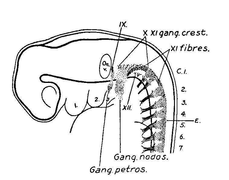
Fig. 2. Reconstruction of peripheral nerves in three weeks human embryo 4.3 mm long
Mall collection No. 148. Enlarged 16.7 diams.
| Historic Disclaimer - information about historic embryology pages |
|---|
 |
- Links: Fig 1 | Fig 2 | Fig 3 | Fig 4 | Fig 5 | Fig 6 | Fig 7 | Fig 8 | Fig 9 | Fig 10 | Fig 11 | Plate 1 | Plate 2 | Plate 3 | Plate 4 | Streeter 1905 | Historic Embryology Papers
Reference
Streeter GL. The Development of the Cranial and Spinal Nerves in the Occipital Region of the Human Embryo. (1905) American J. Anatomy. 4:
Cite this page: Hill, M.A. (2024, April 16) Embryology Streeter1905-fig02.jpg. Retrieved from https://embryology.med.unsw.edu.au/embryology/index.php/File:Streeter1905-fig02.jpg
- © Dr Mark Hill 2024, UNSW Embryology ISBN: 978 0 7334 2609 4 - UNSW CRICOS Provider Code No. 00098G
File history
Click on a date/time to view the file as it appeared at that time.
| Date/Time | Thumbnail | Dimensions | User | Comment | |
|---|---|---|---|---|---|
| current | 20:46, 15 September 2015 | | 758 × 597 (73 KB) | Z8600021 (talk | contribs) | |
| 20:41, 15 September 2015 | 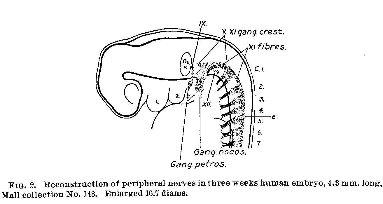 | 1,313 × 699 (107 KB) | Z8600021 (talk | contribs) | {{Streeter1905 figures}} |
You cannot overwrite this file.
File usage
The following page uses this file: