File:Streeter028-30.jpg
Original file (748 × 1,000 pixels, file size: 134 KB, MIME type: image/jpeg)
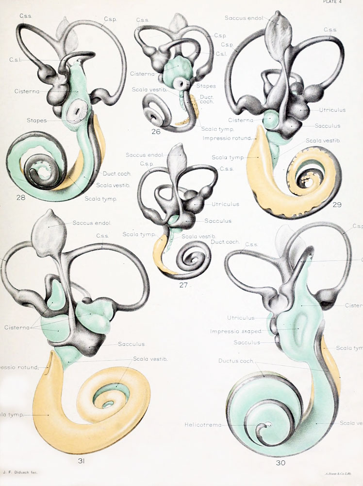
Plate 4
The figures shown on this plate represent a series of median and lateral views of wax-plate reconstructions of the membranous labyrinth and the surrounding periotic tissue-spaces. They illustrate under the same scale of enlargement three typical stages in the development of these spaces.
Abbreviations
- C. s. 1. - ductus semicircuiaris lateralis
- C. s. p. - ductus semicircularis posterior
- C. s. s. - ductus semicircularis superior
- Duct, coch. - ductus cochlearis
- Impressio rotund. - area opposite the fenestra cochlea
- Impressio staped. - area in contact with base of stapes
- Saccus endol. - saccus endolymphaticus
- Scala tymp. - scala tynipani
- Scala vestib. - scala vestibule
Fig. 26
Lateral view of a model reconstructed from a human fetus 50 mm. crown-rump length (Carnegie Collection, No. 84). The cistern and the scala vestibuli are shown in green and the scala tympani is shown in orange. The scala vestibule is in the first stage of its development and consists of a row of large reticular spaces which extend from the ventral margin of the cistern downward along the apical surface of the cochlear duct. The scala tympani is more advanced and shows more complete coalescence of its constituent spaces. Enlarged 11 .4 diameters.
Fig. 27
Median view of the same model shown in figure 26. This view shows the topography of the scala tympani. Its large proximal end lies opposite the fenestra cochlea (rotunda) and corresponds to the focus at which its development originates. Distally it tapers off rapidly where the spaces are smaller and their coalescence less complete. Enlarged 11.4 diameters.
Fig. 28
Lateral view of wax-plate reconstruction of the left membranous labyrinth and the periotic spaces in a human fetus 85 mm. crown-rump length (Carnegie Collection, No. 1400-30), enlarged 11.4 diameters. The cistern and the connecting scala vestibuli are shown in green. Although the greater part of the cistern abuts against the stapes, it will be noted that it is also begiiming to spread over the liorsal surface of the utricle and along the inner border of the lateral semicircular duct. The scala vestibule communicates freely with the cistern and extends downward alotig the apical surface of the cochlear duct, throughout nearly two turns, showing the characteristic sacculated appearance near its tip, where the coalescence of the spaces is less complete.
Fig. 29
Median view of same model shown in figure 28, enlarged 11.4 diameters. The scala tympani is shown in orange. The oval indentation in its proximal end corresponds to the fenestra cochlea (rotunda). This space extends along the cochlear duct about the same distance as the scala vestibuli, but the two do not communicate yet at any place. The peripheral border of the scala tympani is characterized by sacculations corresponding to spaces that are coalescing with the main space. The growth of the scala is due to a coalescence of new spaces along its peripheral border rather than along its central border.
Fig. 30
Lateral view of a wax-plate reconstruction of the left membnmoiis labyrinth and the periotic spaces in a human fetus 130 mm. crown-rump length (Carnegie Collection, No. 1018), enlarged 11.4 diameters. The cistern and scala vestibuli are shown in green and the scala tympani is shown in orange, as in the previous figures. The cartilaginous stapes was removed from this model and the oval impression that it makes on the cistern can be plaiidy seen. The cistern has spread over the top of the utricle and part way along the lateral semicircular duct. The scala vestibuli extends to the ti|) of the cochlear duct, where it communicates with the scala tympani, thas forming the helicotrema.
Fig. 31
Median view of same model shown in figure 30, enlarginl 11.4 diameters. The oval impression on the proximal end of the scala tympani corresponds to the fenestra cochleae (rotunda). As yet there is no conmiunication at this point between the scala tympani and subarachnoid spaces, such :is is found in the adult and known as the aqua-ductus cochleae. The spaces making up the cistern cover almost the whole of the utricle and saccule except the places at which the nerves enter and a small part of the medial surface near the attachment of the appendage.
Reference
Streeter G.L. The histogenesis and growth of the otic capsule and its contained periotic tissue-spaces in the human embryo Contributions to Embryology Carnegie Institution No.20 (1918) pp5-54, 4 text-figures and 4 plates.
File history
Click on a date/time to view the file as it appeared at that time.
| Date/Time | Thumbnail | Dimensions | User | Comment | |
|---|---|---|---|---|---|
| current | 15:24, 22 April 2012 | | 748 × 1,000 (134 KB) | Z8600021 (talk | contribs) | better scan of figures |
| 00:16, 15 February 2011 | 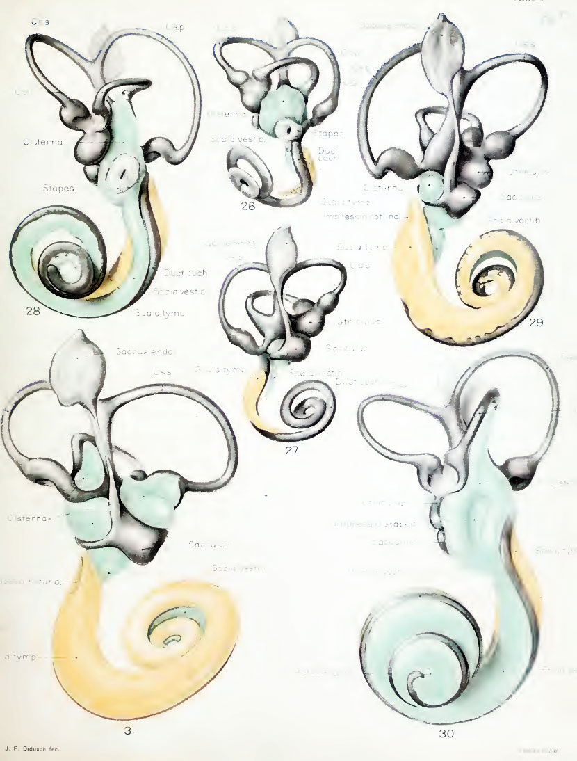 | 830 × 1,094 (143 KB) | S8600021 (talk | contribs) |
You cannot overwrite this file.