File:Streeter014-19.jpg
Original file (640 × 800 pixels, file size: 199 KB, MIME type: image/jpeg)
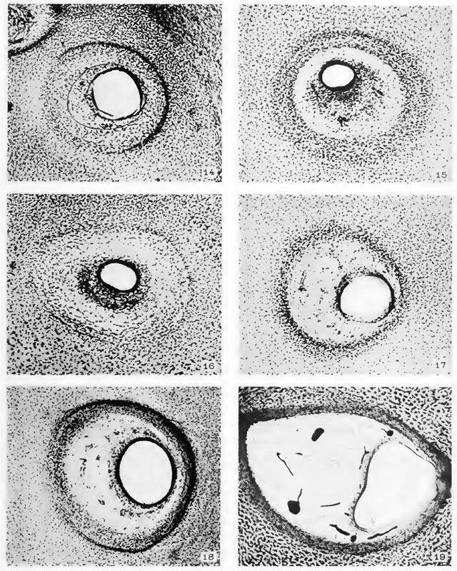
Plate 2
The figures on Plate II are in continuation of those on Plate I and show the final establishment of the periotic reticular tissue. They also show, on being comp.arerl with younger stages, the manner in which the cartilage becomes excavated in order to yield room for the enlarging duct and also to allow for its changing position. The excavation is brought about by the dedifferentiation of cartilage into reticular- tissue. Throughout this period the margin of the cartilaginous canal continues in an unstable condition and is gradually either receding or advancing, through the processes of dedifferentiation, into precartilage or differentiation from precartilage respectively. The periotic reticulum in its later stages develops fibrous membranes at its inner and outer borders. The one at the inner border forms the membrana propria for the epithelial duct, and the one at the outer border becomes the perichondrium.
Fig. 14
Section through the lateral semicircular canal in a human fetu.s 43 mm. crown-rump length (Carnegie Collection, No. 886, slide 42, section 3). The section is 100m thick and is enlarged 90 diameters. The zone of precartilage is expanding around its peripheral margin by dedifferentiation of the surrounding cartilage and on its central margin the precartilage is giving way before the advancing reticulum. A crescentic area of periotic reticulum is established on the median side (to the left) of the epithelial duct, about 8 mm. deep in the photograph.
Fig. 15
Section through the lateral semicircular canal in a human fetus 46 mm. crown-rump length (Carnegie Collection, No. 9.5, slide 72, section 1). The section i.s 100m thick and is enlarged 90 diameters. The original area of precartilage is now all dedifferentiated into reticulum, and a new area of precartilage has formed outside of this at the expense of the surrounding cartilage. The new area of precartilage is about OS cm. deep in the photograph. Everything between this and the epithelium is reticulum, the peripheral part of which is not yet completely vascularized.
Fig. 16
Section thiough the posterior semicircular canal in a human fetus 50 mm. crown-rump length (Carnegie Collection, No. 184, shde 23). The .section is 50^ thick and is enlarged 90 diameters. The dedifferentiation of precartilage into reticulum is nearly complete, there being left ordy a narrow hne of it along the margin of the cartilage. The vascularization of the reticulum is not yet completed. The small diameter and the thick wall of the epithelial duct in this figure and in figure 15 result from contraction. If they were distended in the process of fixation they would doubtless be as large as those in figures 14 and 17.
Fig. 17
Section through the posterior semicircular canal in a human fetus 52 mm. crown-rump length (Carnegie Collection, No. 96, shde 12, section 2). The section is 100m thick and is enlarged 90 diameters. It differs from figure 16 in having a more mature periotic reticulum.
Fig. 18
Section through the posterior semicircular canal in a human fetus, So mm. crown-rump length (Carnegie Collection, No. 1400-30, slide 43, section 2). The section is 100m thick and is enlarged 90 diameters. At the inner margin of the reticulum can now be seen the membrana propria .supporting the semicircular duct and at the outer margin is the thick peri.'hondrium, between which and the cartilage there is a narrow open space that is better seen on the left part of the photograph. The sharp dark line along the margin of the cartilage on the right is an appearance due to the excavation of cartilage at that point. It consists of an intermediate zone in which the cartilage is being dedifferentiated into precartilage and that in turn into reticular tissue.
Fig. 19
Section through the superior semicircular canal in a human fetus 130 mm. crown-rump length (Carnegie Collection, No. 1018, slide 30, section 1). The section is 50m thick and is enlarged 90 diameters. It shows a rather mature perichondrium closely attached to the cartilage, separated from it, however, by a narrow intermediate zone that is not seen in the photograph. This zone is connected with the further enlargement of the cartilaginous canal, the growth of which is not yet completed. In the outer part of the canal the perichondrium fuses with the membrana propria of the semicircular duct. The periotic reticulum is beginning to break up in the formation of larger spaces, which it does by the retraction of its trabecute, thereby allowing adjacent spaces to coalesce. The blood-vessels in this specimen were injected with India ink.
Reference
Streeter G.L. The histogenesis and growth of the otic capsule and its contained periotic tissue-spaces in the human embryo Contributions to Embryology Carnegie Institution No.20 (1918) pp5-54, 4 text-figures and 4 plates.
File history
Click on a date/time to view the file as it appeared at that time.
| Date/Time | Thumbnail | Dimensions | User | Comment | |
|---|---|---|---|---|---|
| current | 00:15, 15 February 2011 | | 640 × 800 (199 KB) | S8600021 (talk | contribs) | |
| 00:14, 15 February 2011 | 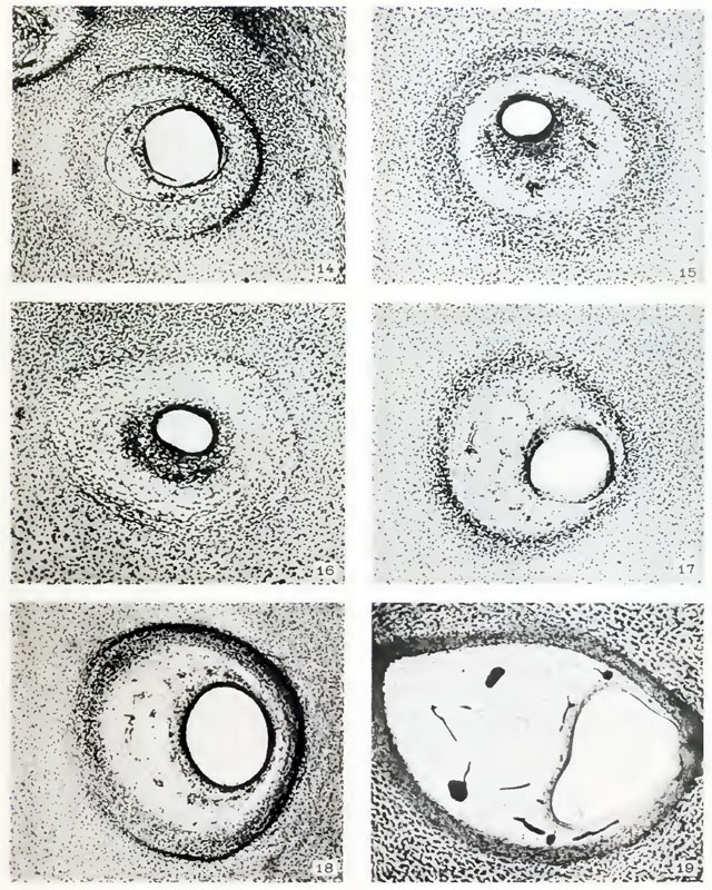 | 640 × 800 (197 KB) | S8600021 (talk | contribs) |
You cannot overwrite this file.
File usage
The following page uses this file: