File:Stewart1955 plate01.jpg
Original file (1,280 × 1,053 pixels, file size: 135 KB, MIME type: image/jpeg)
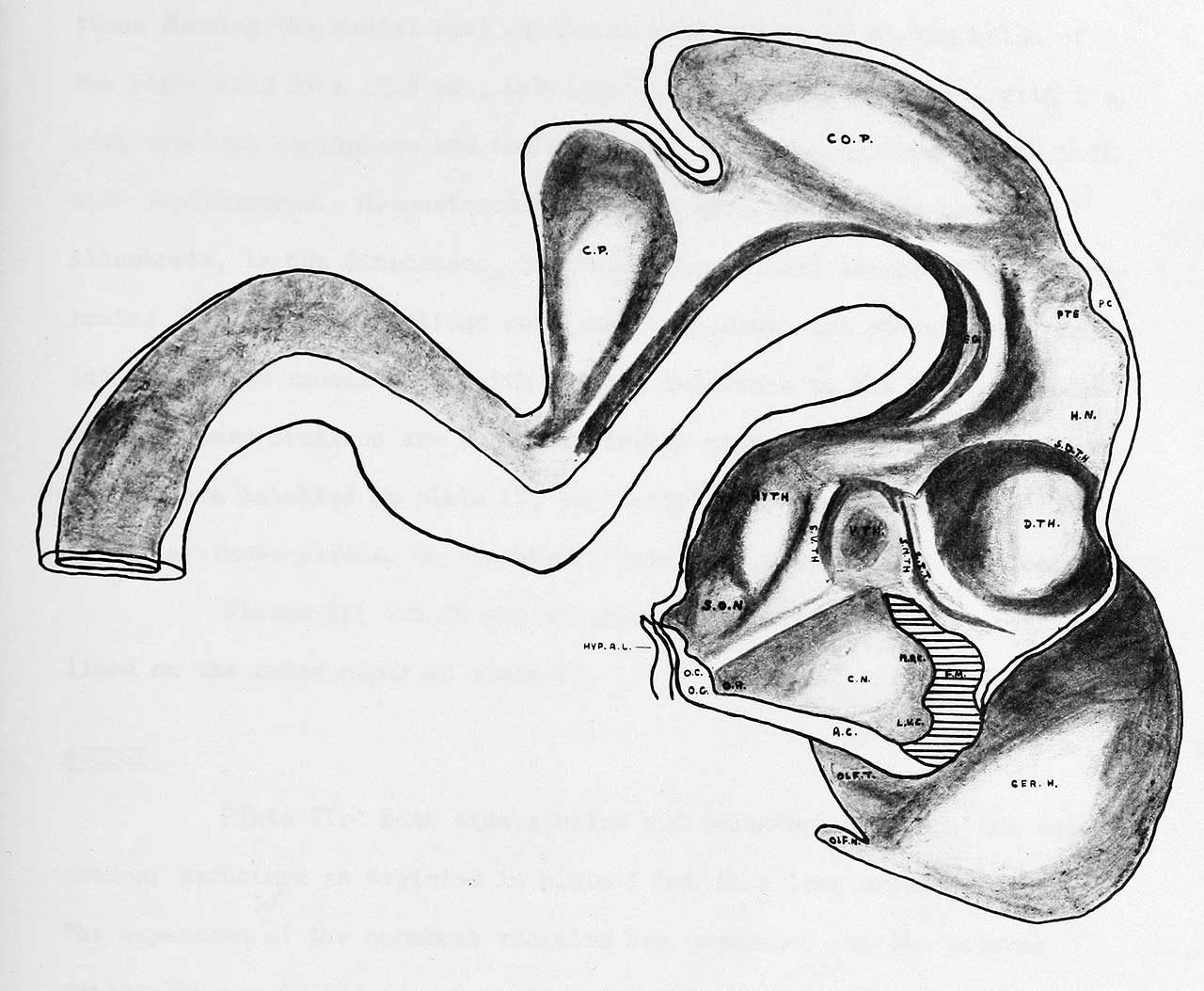
Plate 1 Embryo H51
(original 40X)
This is a photographic reproduction of a graphic reconstruction (a lateral view) of the forebrain of a c C-R length of human Embryo, as outlined in the text.
Represents the left half of the brain of a human embryo of 15.0 mm, C-R length with an estimated ovulation age of 37 days. It is a graphic reconstruction of the configurations of the nuclei forming the medial wall of the mesencephalon and diencephalon and the medial external wall of the anterior portion of the left cerebral hemisphere. This graphic reconstruction was done to provide a basis to which the series of embryos could be related, and was constructed ty serial cross sections. The term nucleus as used in these descriptions does not necessarily indicate a region directly comparable to a nucleus in the adult sense, but indicates an area of cellular concentration which can be identified in the forebrain of successively older embryos. The names given to the tracts and nuclei have been based on the names of corresponding nuclei and tracts in the adult brain wherever it has been possible to identilNr the embryonic structure with its adult derivitive.
Online Editor ovulation age of 37 days is Carnegie stage 16 though 15.5 mm CRL suggests an older age.
| Abbreviations | |
|---|---|
| A.C.A. anterior cerebral artery
A.CH.A. anterior choroidal artery A.CO.A. anterior communicating artery A. COM. anterior commissure A.P. anterior plexus B.A. basilar artery CER.E. cerebral hemisphere C.N. corpus striatum (caudate nucleus) CO.P. collicular plate C.P. cerebellar plate D.A. diencephalic artery D.C.A. diencephalic artery DIM. diencephalon D.TH. dorsal thalamus F. of M. foramen of Monro HIPPO.P. hippocampus primordium H.N. habenular nucleus HYP.A.L. hypophysis anterior lobe HYPOPH. hypophysis HY.TH. hypothalamus I.G. internal capsule I.C.A. Internal carotid artery I.C.V. internal cerebral vein. IT.S. intrathalamic sulcus L.G.B. lateral geniculate boc^r L.S.A. lateral striate arteries M.A. mesencephalic artery M.A.C.C. medial artery of the corpus sallosum M.C.A. middle cerebral artery MES. mesencephalon M.R. mammalary recess M.S.A. medial striate artery (Heubner's recurrent) |
O.A. ophthalmic artery
O.G. optic groove OLF.N. olfactory nerve OLF.TUB. olfactory tubercle Q.N. optic nerve O.R. optic recess O x putamen PA. pallium P.C. posterior commissure PC. A. posterior communicating artery P.CO.A. posterior communicating artery P.CH.A. posterior choroidal artery P.I.C.A. posterior inferior cerebellar artery PIT. pituitary gland primordium P.O.A. primitive olfactory artery PO.R. pre-optic recess PTE. pretecturn RHOMB. rhombencephalon 5.C.A. superior cerebellar artery S.D.TH. dorsal thalamic sulcus S.M.TH. medial thalamic sulcus S.O.N. supra-optic nucleus S.P.C.A. superior portion of the posterior cerebral artery s.s. straight sinus S.S.P. superior sagittal sinus STAPES.A. stapedial artery S.V.TH. ventral thalamic sulcus TEG. tegmentum V.A. vertebral artery VEL. INTER. velum interpositum V.S. ventricular sulcus V.TH. ventral thalamus Z.IT. zona intrathamica |
Reference
Stewart GG. The development of the blood supply to the human embryo basal ganglia. (1955) University of Alberta, Canada.
Cite this page: Hill, M.A. (2024, April 18) Embryology Stewart1955 plate01.jpg. Retrieved from https://embryology.med.unsw.edu.au/embryology/index.php/File:Stewart1955_plate01.jpg
- © Dr Mark Hill 2024, UNSW Embryology ISBN: 978 0 7334 2609 4 - UNSW CRICOS Provider Code No. 00098G
File history
Click on a date/time to view the file as it appeared at that time.
| Date/Time | Thumbnail | Dimensions | User | Comment | |
|---|---|---|---|---|---|
| current | 18:38, 10 June 2018 | | 1,280 × 1,053 (135 KB) | Z8600021 (talk | contribs) | |
| 18:38, 10 June 2018 | 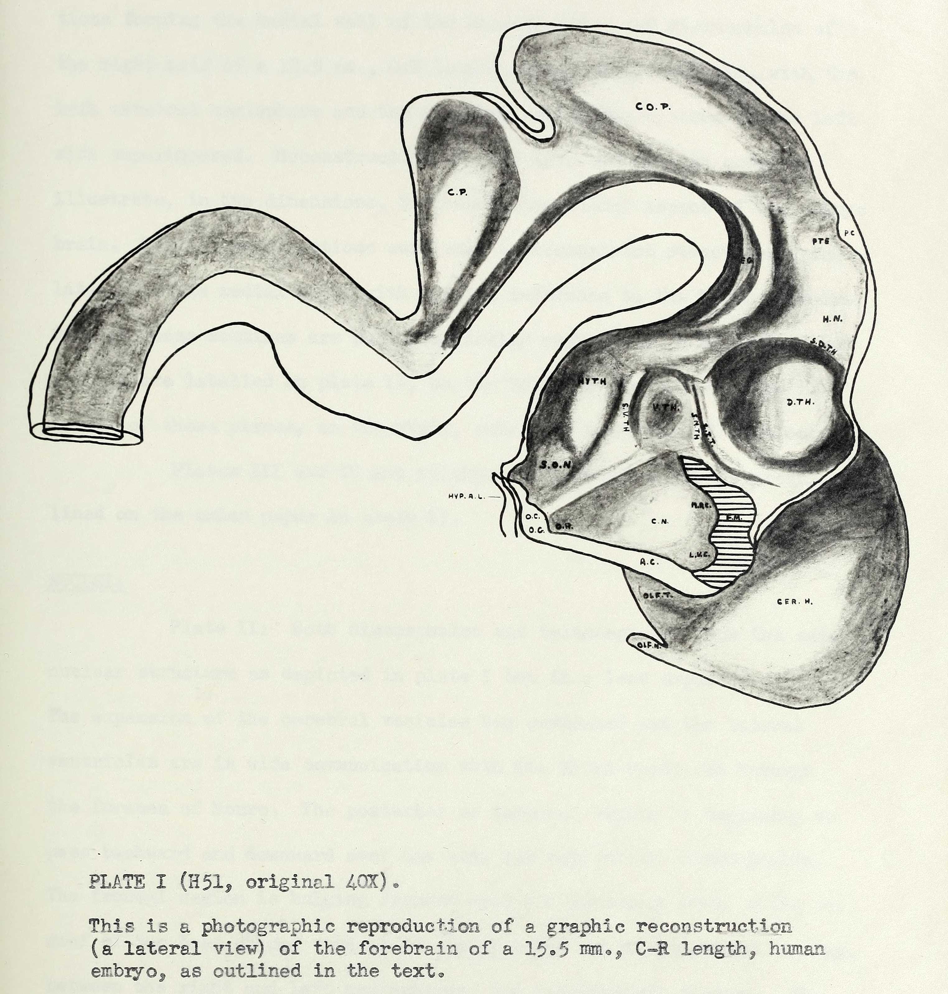 | 3,047 × 3,196 (673 KB) | Z8600021 (talk | contribs) |
You cannot overwrite this file.
File usage
The following page uses this file: