File:Stage 22 image 218.jpg
Original file (1,200 × 730 pixels, file size: 308 KB, MIME type: image/jpeg)
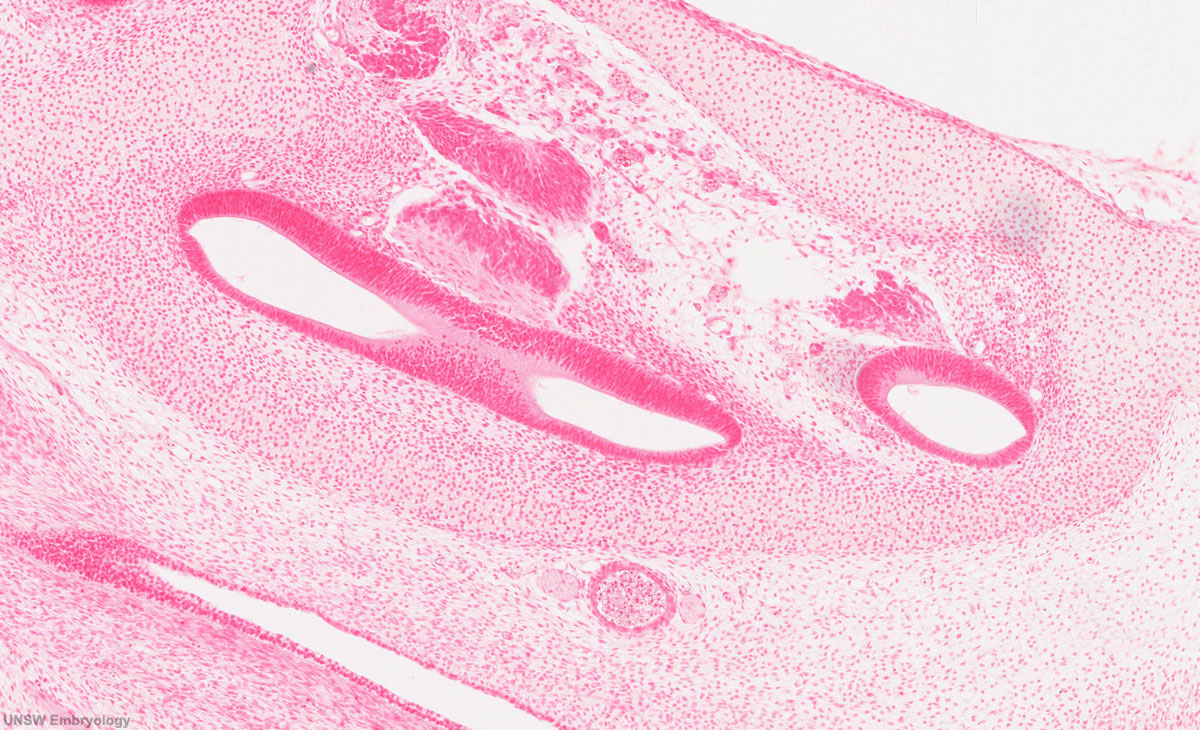
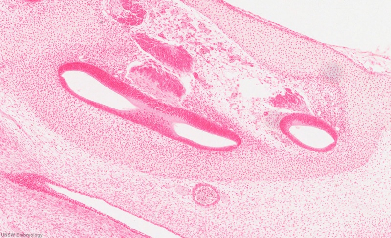
Developing Cochlea
Section through turns in the developing cochlea human embryo week 8 (Carnegie stage 22).
Features
- otic capsule - surrounding the developing inner ear is the cartilage, later to ossify, forming part of the chondrocranium base of the developing skull.
- spiral ganglia - cochlear component of the ganglia (vestibulocochlear nerve, 8th cranial nerve, CN VIII) top of the image.
- internal carotid artery - circular blood vessel lying between developing cochlea and auditory tube.
- auditory tube - cavity at bottom of the image.
| Selected Embryo Histology - Week 8 (Stage 22) |
|---|
|
| Links: Carnegie stage 22 | Week 8 |


Image Source: UNSW Embryology, no reproduction without permission.
Cite this page: Hill, M.A. (2024, April 18) Embryology Stage 22 image 218.jpg. Retrieved from https://embryology.med.unsw.edu.au/embryology/index.php/File:Stage_22_image_218.jpg
- © Dr Mark Hill 2024, UNSW Embryology ISBN: 978 0 7334 2609 4 - UNSW CRICOS Provider Code No. 00098G
File history
Click on a date/time to view the file as it appeared at that time.
| Date/Time | Thumbnail | Dimensions | User | Comment | |
|---|---|---|---|---|---|
| current | 01:35, 23 August 2011 | | 1,200 × 730 (308 KB) | S8600021 (talk | contribs) | Screen-Shot-2011-08-22-at-6.16.32-PM.jpg |
You cannot overwrite this file.
File usage
The following 34 pages use this file:
- BGDA Practical 7 - Week 8
- Carnegie stage 22
- Lecture - Sensory Development
- File:Stage22 vertebra and spinal cord 1.jpg
- File:Stage 22 image 200.jpg
- File:Stage 22 image 201.jpg
- File:Stage 22 image 203.jpg
- File:Stage 22 image 204.jpg
- File:Stage 22 image 205.jpg
- File:Stage 22 image 206.jpg
- File:Stage 22 image 207.jpg
- File:Stage 22 image 208.jpg
- File:Stage 22 image 209.jpg
- File:Stage 22 image 210.jpg
- File:Stage 22 image 211.jpg
- File:Stage 22 image 212.jpg
- File:Stage 22 image 213.jpg
- File:Stage 22 image 214.jpg
- File:Stage 22 image 215.jpg
- File:Stage 22 image 216.jpg
- File:Stage 22 image 217.jpg
- File:Stage 22 image 218.jpg
- File:Stage 22 image 219.jpg
- File:Stage 22 image 220.jpg
- File:Stage 22 image 222.jpg
- File:Stage 22 image 223.jpg
- File:Stage 22 image 224.jpg
- File:Stage 22 image 225.jpg
- File:Stage 22 image 301.jpg
- File:Stage 22 image 302.jpg
- File:Stage 22 image 322.jpg
- File:Stage 22 vomeronasal organ.jpg
- Template:Stage 22 histology gallery
- Template:Stage 22 histology gallery table