File:Stage 22 image 205.jpg

Original file (1,112 × 909 pixels, file size: 261 KB, MIME type: image/jpeg)
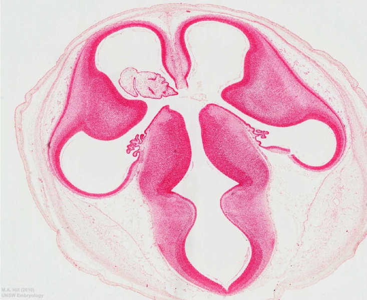
Human Embryo Carnegie stage 22
Ventricular System
- lateral ventricle (anterior and inferior horn)
- interventricular foramen
- third ventricle
- choroid plexus
Neural System
- cortex
- mesencephalic vesicle
- thalamus
- subarachnoid space
In the human embryo identify the large telencephalic vesicles and the choroid plexus. The cavity in these (the lateral ventricles) communicate with the ventricle of the diencephalon (3rd ventricle) through the interventricular foramen. The basal part of the telencephalon forms the basal ganglia, a solid mass. Posteromedially these basal ganglia are in contact with the diencephalon. The large masses in either side of the diencephalon form the thalami.
- Links: Image A3 labeled | neural | ventricular
| Selected Embryo Histology - Week 8 (Stage 22) |
|---|
|
| Links: Carnegie stage 22 | Week 8 |
Cite this page: Hill, M.A. (2024, April 24) Embryology Stage 22 image 205.jpg. Retrieved from https://embryology.med.unsw.edu.au/embryology/index.php/File:Stage_22_image_205.jpg
- © Dr Mark Hill 2024, UNSW Embryology ISBN: 978 0 7334 2609 4 - UNSW CRICOS Provider Code No. 00098G
File history
Click on a date/time to view the file as it appeared at that time.
| Date/Time | Thumbnail | Dimensions | User | Comment | |
|---|---|---|---|---|---|
| current | 17:24, 20 April 2011 |  | 1,112 × 909 (261 KB) | S8600021 (talk | contribs) | ==Human Embryo Carnegie stage 22== Stage 22 image 205.jpg Category:Indexed pages Category:Human Embryo Category:Carnegie Stage Category:Carnegie Stage 22 Category:Week 8 Category:Gastrointestinal Tract [[Category:Musculoskeletal |
You cannot overwrite this file.
File usage
The following 36 pages use this file:
- BGDA Practical 7 - Week 8
- Carnegie stage 22
- Lecture - Neural Development
- Neural - Thalamus Development
- Neural - Ventricular System Development
- File:Stage22 vertebra and spinal cord 1.jpg
- File:Stage 22 image 200.jpg
- File:Stage 22 image 201.jpg
- File:Stage 22 image 203.jpg
- File:Stage 22 image 204.jpg
- File:Stage 22 image 205.jpg
- File:Stage 22 image 206.jpg
- File:Stage 22 image 207.jpg
- File:Stage 22 image 208.jpg
- File:Stage 22 image 209.jpg
- File:Stage 22 image 210.jpg
- File:Stage 22 image 211.jpg
- File:Stage 22 image 212.jpg
- File:Stage 22 image 213.jpg
- File:Stage 22 image 214.jpg
- File:Stage 22 image 215.jpg
- File:Stage 22 image 216.jpg
- File:Stage 22 image 217.jpg
- File:Stage 22 image 218.jpg
- File:Stage 22 image 219.jpg
- File:Stage 22 image 220.jpg
- File:Stage 22 image 222.jpg
- File:Stage 22 image 223.jpg
- File:Stage 22 image 224.jpg
- File:Stage 22 image 225.jpg
- File:Stage 22 image 301.jpg
- File:Stage 22 image 302.jpg
- File:Stage 22 image 322.jpg
- File:Stage 22 vomeronasal organ.jpg
- Template:Stage 22 histology gallery
- Template:Stage 22 histology gallery table


