File:Stage8 sem1.jpg
Original file (828 × 1,000 pixels, file size: 109 KB, MIME type: image/jpeg)
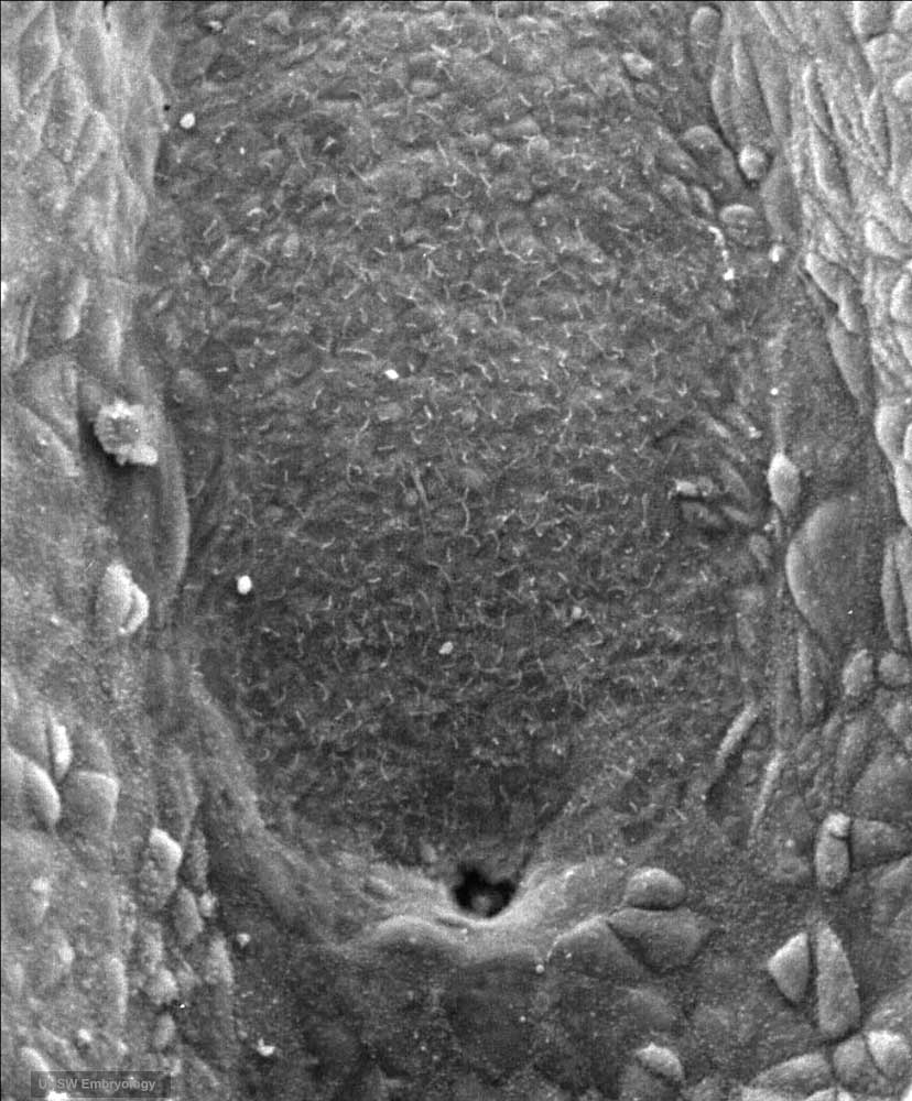
Human Embryo Scanning EM Notochordal Plate
Human embryo (Carnegie stage 8, day 18) SEM image showing the notochordal plate. This is an early embryonic development transient cellular structure and region lying above the primitive streak, that will be converted into the notochord.
- Links: Image - notochordal plate | Image - neurenteric canal | Image - nodal cilia | notochord | Stage 8 | Gastrulation
Image Source: Scanning electron micrographs of the Carnegie stages of the early human embryos are reproduced with the permission of Prof Kathy Sulik, from embryos collected by Dr. Vekemans and Tania Attié-Bitach. Images are for educational purposes only and cannot be reproduced electronically or in writing without permission.
Original file name: Stage8day18notochordalplate.jpg
File history
Click on a date/time to view the file as it appeared at that time.
| Date/Time | Thumbnail | Dimensions | User | Comment | |
|---|---|---|---|---|---|
| current | 08:52, 22 August 2009 | | 828 × 1,000 (109 KB) | S8600021 (talk | contribs) | Stage 8 embryo (day18) showing detail of the notochordal plate Original file name: Stage8day18notochordalplate.jpg {{Template:SEM}} |
You cannot overwrite this file.
File usage
The following 5 pages use this file: