File:Spleen white pulp development model.jpg

Original file (900 × 439 pixels, file size: 59 KB, MIME type: image/jpeg)
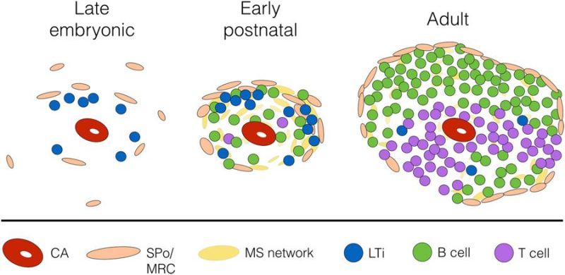
Spleen White Pulp Development Mouse Model
Proposed interactions between spleen organiser cells and LTi/B cells which leads to marginal zone remodelling and adult white pulp formation.
Late embryogenesis (E16.5), CD4+IL-7Rα+ lymphoid tissue inducer (LTi) cells can be observed between the central arteriole (CA) and presumptive MAdCAM-1+VE-Cadherin+ spleen organiser (SPo) cells.
After birth LTi continue to co-localise with SPo (MAdCAM-1+CD31+), which form a primitive marginal zone network surrounding the CD31+ central arterioles. This postnatal developmental stage coincides with B cell migration into the spleen, and both LTi and B cells contribute to lymphotoxin signalling which upregulates MAdCAM-1 expression in the marginal zone, and initiates vascular reorganisation of the Flk-1+ephrinB2+ marginal sinus (MS) network that extends through the white pulp.
Adult MAdCAM-1+CD31+ cells are localised in the marginal zone and represent mature marginal zone reticular cells (MRC).
(figure legend)
Reference
Tan JKH & Watanabe T. (2018). Determinants of postnatal spleen tissue regeneration and organogenesis. NPJ Regen Med , 3, 1. PMID: 29367882 DOI.
Copyright
Open Access This article is licensed under a Creative Commons Attribution 4.0 International License, which permits use, sharing, adaptation, distribution and reproduction in any medium or format, as long as you give appropriate credit to the original author(s) and the source, provide a link to the Creative Commons license, and indicate if changes were made. The images or other third party material in this article are included in the article’s Creative Commons license, unless indicated otherwise in a credit line to the material. If material is not included in the article’s Creative Commons license and your intended use is not permitted by statutory regulation or exceeds the permitted use, you will need to obtain permission directly from the copyright holder. To view a copy of this license, visit http://creativecommons.org/licenses/by/4.0/.
Original file name - 41536_2018_39_Fig1_HTML.jpg
Cite this page: Hill, M.A. (2024, April 19) Embryology Spleen white pulp development model.jpg. Retrieved from https://embryology.med.unsw.edu.au/embryology/index.php/File:Spleen_white_pulp_development_model.jpg
- © Dr Mark Hill 2024, UNSW Embryology ISBN: 978 0 7334 2609 4 - UNSW CRICOS Provider Code No. 00098G
File history
Click on a date/time to view the file as it appeared at that time.
| Date/Time | Thumbnail | Dimensions | User | Comment | |
|---|---|---|---|---|---|
| current | 11:37, 11 February 2019 | | 900 × 439 (59 KB) | Z8600021 (talk | contribs) | 41536_2018_39_Fig1_HTML.jpg |
You cannot overwrite this file.
File usage
The following page uses this file: