File:Spaulding-fig09.jpg

Original file (445 × 607 pixels, file size: 37 KB, MIME type: image/jpeg)
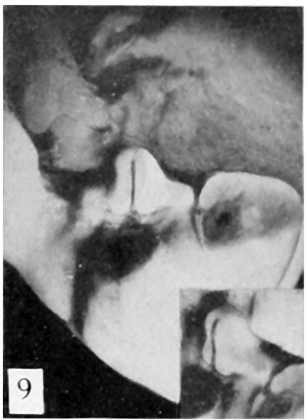
Fig. 9. Carnegie Embryo No. 28
19 mm., male. X 6. (Insert, lateral view.)
Stage 6, 19 mm. CR (fig. 9, male; fig. 13, female). In the male of this stage the phallus is still of narrow, conical form, with the glans area rather more sharply indicated by a broad, band-like depression, not as clearly shown in the photograph as in the embryo itself. The remains of the lateral buttresses arise just proximal to this depression, spreading laterally for a short distance, then continuing basally almost parallel to the axis of the phallus until they finally merge into the tissue at its base. Unlike most of the younger embryos examined, there is almost no caudal trend to these buttresses. The urogenital opening has deepened and become more pronounced. It extends from the base of the phallus practically to its tip. In this specimen it is not limited distally by an epithelial tag. The lack of this appendage, useless as it at present seems, is probably due to some loss prior to or during fixation. The distal portion of the groove (the glans portion) forms a diamond-shaped dilatation extending to the proximal limits of the glans, the remainder of the groove being constricted into a deep, narrow slit. The urethral folds are rather pronounced elevations which diverge distally to merge into the glans, while proximally they are considerably thickened into a pair of caudally projecting, diverging masses (bulbo-urethral swellings?) which merge laterally into the outhing labio-scrotal swellings. They are separated from the cavernous portion of the phallus by broad concavities. Perhaps the most interesting feature of this embryo is that it shows the apparent beginnings of the labio-scrotal swellings. These are a pair of flat, elevated areas on either side of the base of the phallus with which they seem to be continuous. The cranial margins of these areas are nearly straight, extending at diverging angles into the inguinal regions, where their cranio-lateral angles continue for a short distance as ridges of tissue. The caudal margins appear as continuations of the diverging wings of the basal portions of the urethral folds, forming sinuous curves to the caudo-lateral angles, from which points they sweep cranially in rounding curves to the cranio-lateral angles, where they unite with the cranial margins. This is the only specimen which shows these swellings definitely connected with the base of the phallus. In all others in which they are developed there is a groove (lateral phallic groove) on each side which separates them from the base of the phallus.
- Figure Links: Text | Text Figure 1 | Text Figure 2 | Plate 1 | Fig. 1 | Fig. 2 | Fig. 3 | Fig. 4 | Fig. 5 | Fig. 6 | Plate 2 | Fig. 7 | Fig. 8 | Fig. 9 | Fig. 10 | Fig. 11 | Fig. 12 | Fig. 13 | Fig. 14 | Fig. 15 | Fig. 16 | Fig. 17 | Fig. 18 | Fig. 19 | Fig. 20 | Fig. 21 | Fig. 22 | Plate 3 | Fig. 23 | Fig. 24 | Fig. 25 | Fig. 26 | Fig. 27 | Fig. 28 | Fig. 29 | Plate 4 | Fig. 30 | Fig. 31 | Fig. 32 |Fig. 33 | Fig. 34 | Fig. 35 | Fig. 36 | Fig. 37 | Fig. 38 | Fig. 39 | Fig. 40 | Fig. 41 | Fig. 42 | Fig. 43 | Fig. 44 | Fig. 45 | Fig. 46 | Fig. 47 | Fig. 48 | Fig. 49 | Fig. 50 | Fig. 51 | Fig. 52 | Fig. 53 | Fig. 54
| Historic Disclaimer - information about historic embryology pages |
|---|
| Pages where the terms "Historic" (textbooks, papers, people, recommendations) appear on this site, and sections within pages where this disclaimer appears, indicate that the content and scientific understanding are specific to the time of publication. This means that while some scientific descriptions are still accurate, the terminology and interpretation of the developmental mechanisms reflect the understanding at the time of original publication and those of the preceding periods, these terms, interpretations and recommendations may not reflect our current scientific understanding. (More? Embryology History | Historic Embryology Papers) |
Reference
Spaulding MH. The development of the external genitalia in the human embryo. (1921) Contrib. Embryol., Carnegie Inst. Wash. Publ. 81, 13: 69 – 88.
Cite this page: Hill, M.A. (2024, April 19) Embryology Spaulding-fig09.jpg. Retrieved from https://embryology.med.unsw.edu.au/embryology/index.php/File:Spaulding-fig09.jpg
- © Dr Mark Hill 2024, UNSW Embryology ISBN: 978 0 7334 2609 4 - UNSW CRICOS Provider Code No. 00098G
File history
Click on a date/time to view the file as it appeared at that time.
| Date/Time | Thumbnail | Dimensions | User | Comment | |
|---|---|---|---|---|---|
| current | 00:15, 15 April 2015 |  | 445 × 607 (37 KB) | Z8600021 (talk | contribs) |
You cannot overwrite this file.
File usage
The following 3 pages use this file:
