File:Spaulding-fig04.jpg

Original file (933 × 750 pixels, file size: 110 KB, MIME type: image/jpeg)
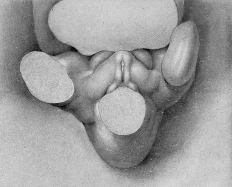
Fig. 4. Carnegie Embryo 1784a
12 mm X 21.
Stage 3, 8 to 12 mm. (fig. 3 and fig. 4). As growth proceeds, the genital tubercle is transformed into a compressed, conical protuberance. This is brought about by the deepening of the umbihco-phallic groove, so that the cranial slope of the tubercle is nearly straight (i. e., approximately at right angles with the body axis), while the caudal slope remains decidedly convex. At the same time the caudal outline has become markedly triangular by the broadening of the base until it occupies practically the entire area between the bases of the legs. The conspicuous lateral slopes form the "lateral buttresses" which, arising from the cranial border of the tubercle just proximal to its apical area, have a decidedly caudal trend, so that they finally disappear basally opposite the caudal border of the tubercle. The apex of the tubercle is now clearly marked off from the more proximal portion by a shallow circular depression, indicating it as the future glans and separating it from the basal shaft. The urethral groove is a long, lancet-shaped depression, broadest and deepest basally, narrowing distally into a shallow slit limited by a very small "epithelial tag" just proximal to the primitive glans area. The urethral folds (margins of the groove) are elevated as shght rolls of tissue which distally merge into the glans region, while basally they become more tumid and broaden out to surround the anal pit as anal tubercles. Lateral to these urethral folds, the caudal surface of the tubercle is somewhat swollen in the younger specimen illustrating this stage (fig. 3), in marked contrast to the decided concavity of these regions in the older embryo (fig. 4) . In the former (fig. 3) the urethral and anal membranes have not ruptured. The older embryo (fig. 4) agrees with the finding that (in the majority of cases) these membranes rupture at about the stage of 12 to 13 mm., although several specimens were found, among both the sectioned and unsectioned material in which the membranes were still imperforate at 17 mm. The perforation of this membrane transforms the shallow urethral groove into a gutter-like, primitive urogenital opening as a direct communication between the phallic portion of the urogenital sinus and the exterior. This is accompanied by an increase in the definiteness of its outlines, as the result of which its sex difference in length is correspondingly emphasized. Because of the variation in the time of rupturing of this membrane, as well as on account of normal differences in the breadth of the opening thus formed, it seems advisable to continue to use the term urethral groove, except when referring to embryos that clearly show this feature as an opening.
| Historic Disclaimer - information about historic embryology pages |
|---|
| Pages where the terms "Historic" (textbooks, papers, people, recommendations) appear on this site, and sections within pages where this disclaimer appears, indicate that the content and scientific understanding are specific to the time of publication. This means that while some scientific descriptions are still accurate, the terminology and interpretation of the developmental mechanisms reflect the understanding at the time of original publication and those of the preceding periods, these terms, interpretations and recommendations may not reflect our current scientific understanding. (More? Embryology History | Historic Embryology Papers) |
- Figure Links: Text | Text Figure 1 | Text Figure 2 | Plate 1 | Fig. 1 | Fig. 2 | Fig. 3 | Fig. 4 | Fig. 5 | Fig. 6 | Plate 2 | Fig. 7 | Fig. 8 | Fig. 9 | Fig. 10 | Fig. 11 | Fig. 12 | Fig. 13 | Fig. 14 | Fig. 15 | Fig. 16 | Fig. 17 | Fig. 18 | Fig. 19 | Fig. 20 | Fig. 21 | Fig. 22 | Plate 3 | Fig. 23 | Fig. 24 | Fig. 25 | Fig. 26 | Fig. 27 | Fig. 28 | Fig. 29 | Plate 4 | Fig. 30 | Fig. 31 | Fig. 32 |Fig. 33 | Fig. 34 | Fig. 35 | Fig. 36 | Fig. 37 | Fig. 38 | Fig. 39 | Fig. 40 | Fig. 41 | Fig. 42 | Fig. 43 | Fig. 44 | Fig. 45 | Fig. 46 | Fig. 47 | Fig. 48 | Fig. 49 | Fig. 50 | Fig. 51 | Fig. 52 | Fig. 53 | Fig. 54
| Historic Disclaimer - information about historic embryology pages |
|---|
| Pages where the terms "Historic" (textbooks, papers, people, recommendations) appear on this site, and sections within pages where this disclaimer appears, indicate that the content and scientific understanding are specific to the time of publication. This means that while some scientific descriptions are still accurate, the terminology and interpretation of the developmental mechanisms reflect the understanding at the time of original publication and those of the preceding periods, these terms, interpretations and recommendations may not reflect our current scientific understanding. (More? Embryology History | Historic Embryology Papers) |
Reference
Spaulding MH. The development of the external genitalia in the human embryo. (1921) Contrib. Embryol., Carnegie Inst. Wash. Publ. 81, 13: 69 – 88.
Cite this page: Hill, M.A. (2024, April 20) Embryology Spaulding-fig04.jpg. Retrieved from https://embryology.med.unsw.edu.au/embryology/index.php/File:Spaulding-fig04.jpg
- © Dr Mark Hill 2024, UNSW Embryology ISBN: 978 0 7334 2609 4 - UNSW CRICOS Provider Code No. 00098G
File history
Click on a date/time to view the file as it appeared at that time.
| Date/Time | Thumbnail | Dimensions | User | Comment | |
|---|---|---|---|---|---|
| current | 23:22, 14 April 2015 |  | 933 × 750 (110 KB) | Z8600021 (talk | contribs) |
You cannot overwrite this file.
File usage
The following 2 pages use this file:
