File:Somite cartoon1.png
Somite_cartoon1.png (400 × 300 pixels, file size: 9 KB, MIME type: image/png)
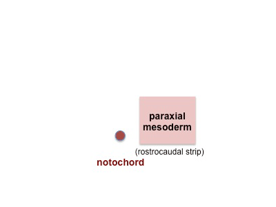
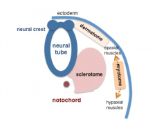
1. Somite Development - Paraxial Mesoderm
Mesoderm lying right and left beside the notochord (axial mesoderm) forms the paraxial mesoderm as a pair of strips along the rostro-caudal axis of the embryo. This paraxial mesoderm will then commence to segment in pairs in a rostro-caudal sequence, the paraxial mesoderm in the head region will remain unsegmented.
Note - the cartoons show just the embryo righthand side mesoderm development (the same events occur on the lefthand side).
- Somite Links: 1 paraxial | 2 early somite | 3 sclerotome and dermomyotome | 4 dermatome and myotome | 5 somite spreading | SEM image - Human Embryo (week 4) showing somites | Movie - somitogenesis Hes expression
- Somite Cartoons

paraxial
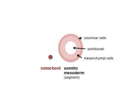
early somite
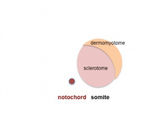
sclerotome and dermomyotome
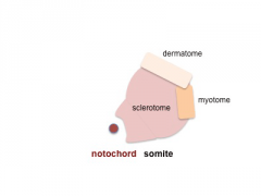
dermatome and myotome
somite spreading


Cite this page: Hill, M.A. (2024, April 18) Embryology Somite cartoon1.png. Retrieved from https://embryology.med.unsw.edu.au/embryology/index.php/File:Somite_cartoon1.png
- © Dr Mark Hill 2024, UNSW Embryology ISBN: 978 0 7334 2609 4 - UNSW CRICOS Provider Code No. 00098G
File history
Click on a date/time to view the file as it appeared at that time.
| Date/Time | Thumbnail | Dimensions | User | Comment | |
|---|---|---|---|---|---|
| current | 18:00, 16 May 2014 | | 400 × 300 (9 KB) | Z8600021 (talk | contribs) | |
| 10:40, 10 August 2009 | 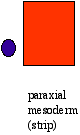 | 80 × 135 (992 bytes) | MarkHill (talk | contribs) | Somite Development cartoon Mesoderm beside the notochord (axial mesoderm) thickens, forming the paraxial mesoderm as a pair of strips along the rostro-caudal axis. Image source: UNSW Embryology http://embryology.med.unsw.edu.au/Notes/skmus.htm#Somite1 |
You cannot overwrite this file.
File usage
The following 30 pages use this file:
- 2009 Lecture 5
- 2010 BGD Lecture - Development of the Embryo/Fetus 1
- 2010 BGD Lecture - Development of the Embryo/Fetus 2
- 2010 BGD Practical 6 - Week 3
- 2010 Lab 3
- 2010 Lecture 5
- 2011 Lab 3 - Week 3
- ANAT2341 Lab 3 - Week 3
- BGDA Lecture - Development of the Embryo/Fetus 1
- BGDA Lecture - Development of the Embryo/Fetus 2
- BGDA Practical 7 - Week 3
- Developmental Mechanism - Epithelial Mesenchymal Transition
- Lecture - Mesoderm Development
- Musculoskeletal System - Limb Development
- Musculoskeletal System Development
- Somite Musculoskeletal Movie
- Somitogenesis
- Talk:2010 BGD Practical 6 - Week 3
- Talk:2011 Lab 3
- File:Mesoderm cartoon 05.jpg
- File:Mesoderm cartoon 06.jpg
- File:Mesoderm cartoon 07.jpg
- File:Mesoderm cartoon 08.jpg
- File:Mesoderm cartoon 09.jpg
- File:Somite cartoon1.png
- File:Somite cartoon2.png
- File:Somite cartoon3.png
- File:Somite cartoon4.png
- File:Somite cartoon5.png
- Template:Somite cartoon