File:Smellie1754 table 02.jpg
From Embryology
Size of this preview: 439 × 600 pixels. Other resolution: 992 × 1,355 pixels.
Original file (992 × 1,355 pixels, file size: 395 KB, MIME type: image/jpeg)
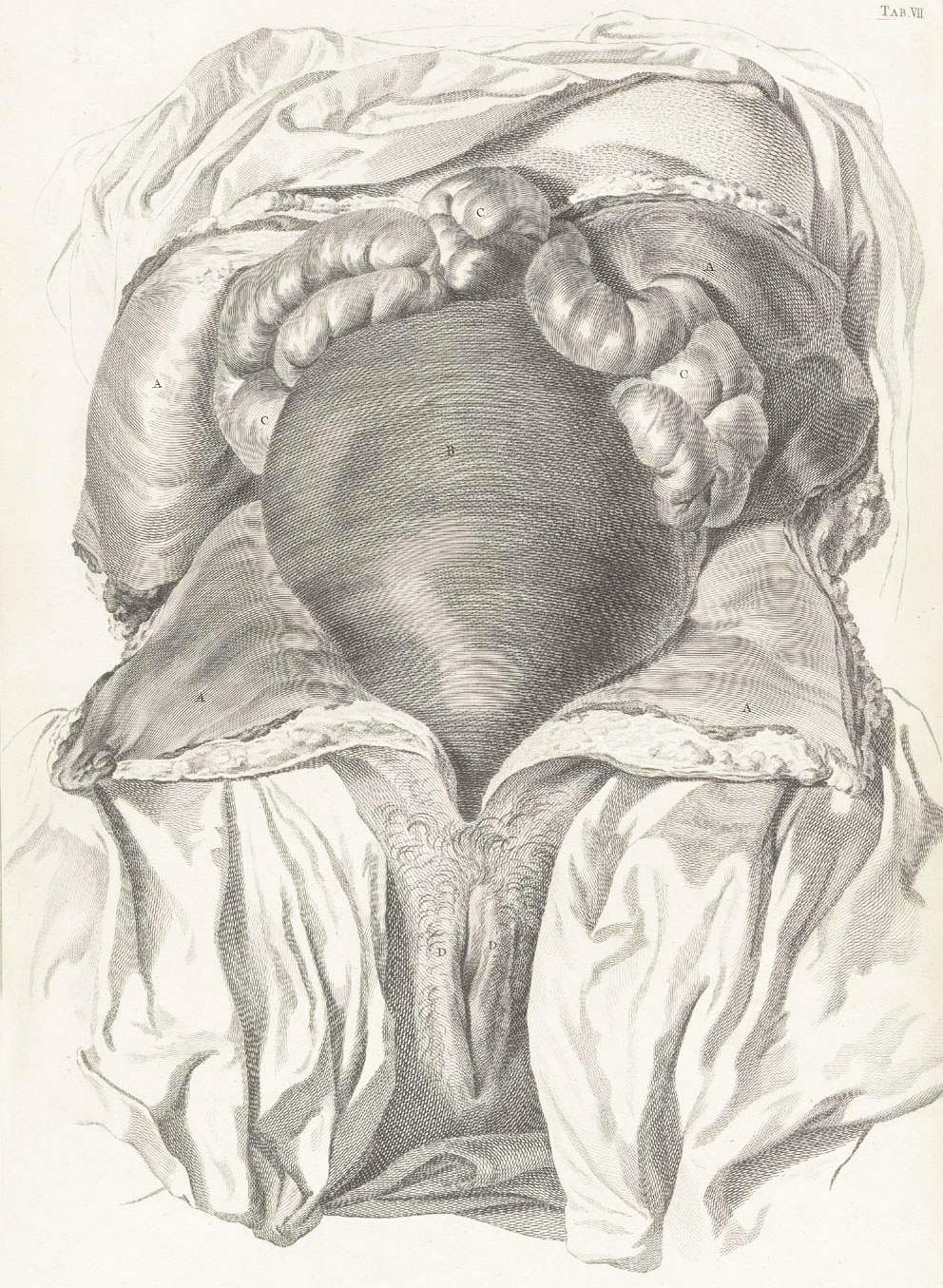
Table 2 Gives a lateral and internal view of the Pelvis divided longitudinally
| This Plate shews the distance from the superior part of the os sacrum to the ossa pubis, as well as from the last-mentioned bones to the coccyx, which in each amounts to about four inches and a quarter. The depth likewise of the posterior, lateral, and anterior parts of the pelvis, is shewn, not in the line of the body, but in that of the pelvis from its brim downward, which is generally three times deeper on the posterior than anterior part, and twice the depth of the last at the sides.
From this view appears also the angle which is formed by the last vertebra of the loins and the superior part of the os sacrum, as likewise the concavity or hollow space in the posterior internal part of the pelvis, arising from the curvature of the last-mentioned bone and coccyx ; finally, the distance from which to the posterior parts of the ossa ischium is here expressed. Vide Tab. XVI. XVII. XVIII. XIX. Also Vol. I. and II as referred to in the former Table. |
Legend
|
| Historic Disclaimer - information about historic embryology pages |
|---|
 |
File history
Click on a date/time to view the file as it appeared at that time.
| Date/Time | Thumbnail | Dimensions | User | Comment | |
|---|---|---|---|---|---|
| current | 12:05, 12 November 2012 | | 992 × 1,355 (395 KB) | Z8600021 (talk | contribs) |
You cannot overwrite this file.
File usage
The following file is a duplicate of this file (more details):
The following 2 pages use this file: