File:Skull CT normal sutures 03.jpg
Original file (600 × 800 pixels, file size: 63 KB, MIME type: image/jpeg)
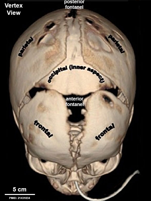
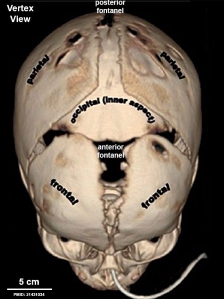
Newborn Skull (CT, vertex view) Showing Anterior and Posterior Fontanels
Computed Tomography (CT) scan with 3D surface-rendered reconstruction of vertex view newborn skull with fontanels (fontanelles) and sutures. Cranial vault bones usually ossify from the center to periphery, which results in this “widened” appearance of the sutures in the newborn.
- Anterior Fontanel (bregmatic fontanelle) - largest fontanel located at the junction of the sagittal, coronal, and frontal sutures. In the newborn it is lozenge-shaped and measures about 4 cm in its antero-posterior and 2.5 cm in its transverse diameter.
- Posterior Fontanel - located at the junction of the sagittal and lambdoidal sutures and is triangular-shaped.
- Lateral Fontanels - laterally located and correspond respectively with the sphenoidal and mastoid angles of the parietal bones, and are small irregular shaped.
- Skull CT Images: Normal overview | Normal vertex and lateral | Normal endocranial and vertex | Normal Vertex - Fontanels | Dolichocephaly and Scaphocephaly | Coronal Synostosis | Anterior Plagiocephaly | Turricephaly | Posterior Plagiocephaly | Deformational Plagiocepahly | Trigonocephaly | Oxycephaly | Computed Tomography
- Skull Links: Skull Development | Historic - skull of a human fetus of 43 millimeters greatest length | Computed Tomography
Reference
Khanna PC, Thapa MM, Iyer RS & Prasad SS. (2011). Pictorial essay: The many faces of craniosynostosis. Indian J Radiol Imaging , 21, 49-56. PMID: 21431034 DOI.
Copyright
Paritosh C Khanna © 2007 - 2012 Indian Journal of Radiology and Imaging
This is an open-access article distributed under the terms of the Creative Commons Attribution License, which permits unrestricted use, distribution, and reproduction in any medium, provided the original work is properly cited. Attribution-NonCommercial-ShareAlike 3.0 Unported (CC BY-NC-SA 3.0)
Original file name: Figure 1(A): IJRI-21-49-g001.jpg http://www.ijri.org/viewimage.asp?img=IndianJRadiolImaging_2011_21_1_49_76055_f2.jpg resized and relabelled.
Cite this page: Hill, M.A. (2024, April 24) Embryology Skull CT normal sutures 03.jpg. Retrieved from https://embryology.med.unsw.edu.au/embryology/index.php/File:Skull_CT_normal_sutures_03.jpg
- © Dr Mark Hill 2024, UNSW Embryology ISBN: 978 0 7334 2609 4 - UNSW CRICOS Provider Code No. 00098G
File history
Click on a date/time to view the file as it appeared at that time.
| Date/Time | Thumbnail | Dimensions | User | Comment | |
|---|---|---|---|---|---|
| current | 10:16, 22 March 2016 | | 600 × 800 (63 KB) | Z8600021 (talk | contribs) | ==Newborn Skull (CT, vertex view) Showing Anterior and Posterior Fontanels== Computed Tomography (CT) scan with 3D surface-rendered reconstruction of vertex view newborn skull with fontanels and sutures. Cranial vault bones usually ossify from the cen... |
You cannot overwrite this file.
File usage
The following 5 pages use this file: