File:Skull CT abnormal 04.jpg
Original file (1,000 × 646 pixels, file size: 102 KB, MIME type: image/jpeg)
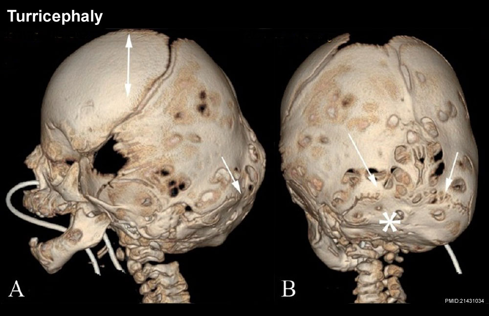
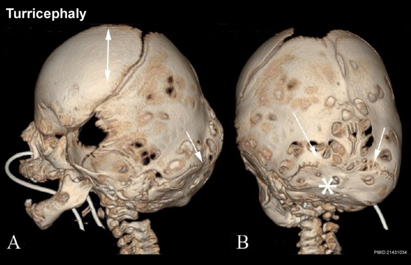
Skull Turricephaly
- arrows - turricephaly secondary to bilateral lambdoid fusion.
- asterisk - small underdeveloped posterior fossa.
- double-headed arrow - "tall" cranium.
- Skull CT Images: Normal overview | Normal vertex and lateral | Normal endocranial and vertex | Normal Vertex - Fontanels | Dolichocephaly and Scaphocephaly | Coronal Synostosis | Anterior Plagiocephaly | Turricephaly | Posterior Plagiocephaly | Deformational Plagiocepahly | Trigonocephaly | Oxycephaly | Computed Tomography
Views
- A - Lateral 3DCT volume rendered image
- B - posterior 3DCT volume rendered image
Reference
Khanna PC, Thapa MM, Iyer RS & Prasad SS. (2011). Pictorial essay: The many faces of craniosynostosis. Indian J Radiol Imaging , 21, 49-56. PMID: 21431034 DOI.
Copyright
This is an open-access article distributed under the terms of the Creative Commons Attribution License, which permits unrestricted use, distribution, and reproduction in any medium, provided the original work is properly cited.
Paritosh C Khanna © 2007 - 2012 Indian Journal of Radiology and Imaging
Attribution-NonCommercial-ShareAlike 3.0 Unported (CC BY-NC-SA 3.0)
Original file name: Figure 5 (A,B) Original figure has been modified, resized and relabelled.
http://www.ijri.org/viewimage.asp?img=IndianJRadiolImaging_2011_21_1_49_76055_f6.jpg
Cite this page: Hill, M.A. (2024, April 19) Embryology Skull CT abnormal 04.jpg. Retrieved from https://embryology.med.unsw.edu.au/embryology/index.php/File:Skull_CT_abnormal_04.jpg
- © Dr Mark Hill 2024, UNSW Embryology ISBN: 978 0 7334 2609 4 - UNSW CRICOS Provider Code No. 00098G
File history
Click on a date/time to view the file as it appeared at that time.
| Date/Time | Thumbnail | Dimensions | User | Comment | |
|---|---|---|---|---|---|
| current | 11:11, 17 March 2012 | | 1,000 × 646 (102 KB) | Z8600021 (talk | contribs) |
You cannot overwrite this file.
File usage
The following 4 pages use this file: