File:Simkins1928 plate05.jpg
From Embryology

Size of this preview: 364 × 600 pixels. Other resolution: 1,281 × 2,111 pixels.
Original file (1,281 × 2,111 pixels, file size: 154 KB, MIME type: image/jpeg)
21 Cross—section of the testis of an embryo of 54 mm. X 200.
22 Detail of the large and small cells in the ovary of a 51-mm. embryo. X 1500.
| Historic Disclaimer - information about historic embryology pages |
|---|
 |
- Simkins 1928: plate 1 | plate 2 | plate 3 | plate 4 | plate 5 | plate 6 | plate 7 | plate 8 | plate 9 | plate 10
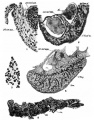
plate 1
plate 2
plate 3
plate 4
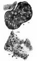
plate 5
plate 6
plate 7
plate 8
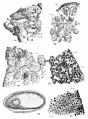
plate 9
plate 10


Reference
Simkins CS. Origin of the sex cells in man. (1928) Amer. J Anat. 41: 248-272.
Cite this page: Hill, M.A. (2024, April 24) Embryology Simkins1928 plate05.jpg. Retrieved from https://embryology.med.unsw.edu.au/embryology/index.php/File:Simkins1928_plate05.jpg
- © Dr Mark Hill 2024, UNSW Embryology ISBN: 978 0 7334 2609 4 - UNSW CRICOS Provider Code No. 00098G
File history
Click on a date/time to view the file as it appeared at that time.
| Date/Time | Thumbnail | Dimensions | User | Comment | |
|---|---|---|---|---|---|
| current | 16:50, 31 January 2018 | | 1,281 × 2,111 (154 KB) | Z8600021 (talk | contribs) | |
| 16:22, 31 January 2018 |  | 1,566 × 2,319 (139 KB) | Z8600021 (talk | contribs) | {{Simkins1928 figures}} |
You cannot overwrite this file.
File usage
The following 12 pages use this file:
- Paper - Origin of the sex cells in man
- File:Simkins1928 plate01.jpg
- File:Simkins1928 plate02.jpg
- File:Simkins1928 plate03.jpg
- File:Simkins1928 plate04.jpg
- File:Simkins1928 plate05.jpg
- File:Simkins1928 plate06.jpg
- File:Simkins1928 plate07.jpg
- File:Simkins1928 plate08.jpg
- File:Simkins1928 plate09.jpg
- File:Simkins1928 plate10.jpg
- Template:Simkins1928 figures