File:Secondary Oocyte during in-vitro fertilisation.jpeg
From Embryology
No higher resolution available.
Secondary_Oocyte_during_in-vitro_fertilisation.jpeg (800 × 558 pixels, file size: 48 KB, MIME type: image/jpeg)
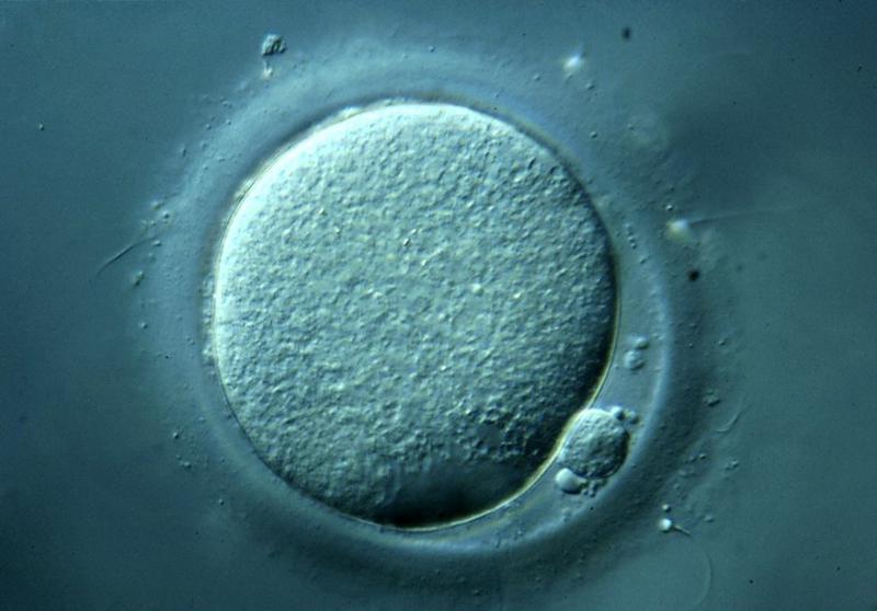
A human secondary oocyte during in-vitro fertilisation with a polar body, viewed with a light microscope.
Reference
S. Walker, Wellcome Images, secondary oocyte during in-vitro fertilisation [1]
Copyright
This is a non-commercial, no derivatives licence distributed under the terms of the Creative Commons Attribution licence [2] which permits restricted use, distribution, and reproduction, provided the original work is properly cited and not modified.
File history
Click on a date/time to view the file as it appeared at that time.
| Date/Time | Thumbnail | Dimensions | User | Comment | |
|---|---|---|---|---|---|
| current | 21:48, 20 August 2014 | | 800 × 558 (48 KB) | Z3418698 (talk | contribs) | A human secondary oocyte during in-vitro fertilisation viewed with a light microscope. The small cell at the bottom right is a polar body. Spice Walker, Wellcome Images. z3418698 |
You cannot overwrite this file.
File usage
There are no pages that use this file.