File:Sea urchin SEM02.jpg
From Embryology

Size of this preview: 800 × 570 pixels. Other resolution: 1,000 × 712 pixels.
Original file (1,000 × 712 pixels, file size: 120 KB, MIME type: image/jpeg)
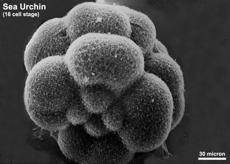
Sea Urchin
Scanning electron microscope image of Lytechinus pictus [sea urchin] embryo at the 16-cell stage.
- four large macromeres are behind the four small micromeres.
- eight mesomeres are behind the macromeres.
- remnants of the fertilization envelope can be seen attached to some of the cells.
Class Echinoidea - Superorder Echinacea -Order Temnopleuroida - Lytechinus pictus
- Links: Sea Urchin Development
JEOL 35C SEM Evelyn Spiegel, Louisa Howard
Image Source: Dartmouth Electron Microscope Facility A variety of scanning and transmission electron microscope images (These images are in the public domain)
(original image scaled, contrast altered and background tidied)
File history
Click on a date/time to view the file as it appeared at that time.
| Date/Time | Thumbnail | Dimensions | User | Comment | |
|---|---|---|---|---|---|
| current | 14:49, 1 June 2011 | | 1,000 × 712 (120 KB) | S8600021 (talk | contribs) | ==Sea Urchin== Scanning electron microscope image of Lytechinus pictus [sea urchin] embryo at the 16-cell stage. * four large macromeres are behind the four small micromeres. * eight mesomeres are behind the macromeres. * remnants of the fertilization |
You cannot overwrite this file.
File usage
The following 2 pages use this file: