File:Screen Shot 2017-10-16 at 1.43.55 pm.png

Original file (1,183 × 535 pixels, file size: 704 KB, MIME type: image/png)
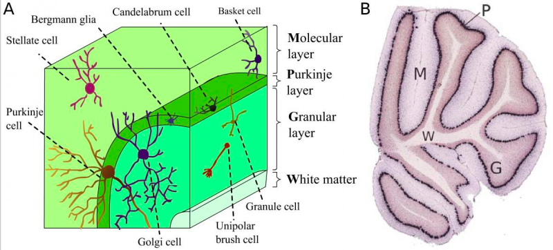
Cortical Layers
(A). Cell types and where they are found in the cerebellar cortical layers.
(B) Shows how the different layers of the cerebellum can be easily determined.
P - the Purkinje layer; G - the granular layer; M - the molecular layer; W - the white matter.
Reference
Abbasi A, Voelter W & Zaidi ZH. (1986). Isolation purification and properties of a site-specific proteolytic enzyme "valyl-proteinase" from Candida tropicalis. Biol. Chem. Hoppe-Seyler , 367, 441-5. PMID: 3527225
Copyright
This is an open-access article distributed under the terms of the Creative Commons Attribution License, which permits unrestricted use, distribution, and reproduction in any medium, provided the original author and source are properly credited.
- Note - This image was originally uploaded as part of an undergraduate science student project and may contain inaccuracies in either description or acknowledgements. Students have been advised in writing concerning the reuse of content and may accidentally have misunderstood the original terms of use. If image reuse on this non-commercial educational site infringes your existing copyright, please contact the site editor for immediate removal.
File history
Click on a date/time to view the file as it appeared at that time.
| Date/Time | Thumbnail | Dimensions | User | Comment | |
|---|---|---|---|---|---|
| current | 12:52, 16 October 2017 | | 1,183 × 535 (704 KB) | Z5076158 (talk | contribs) | (A). Cell types and where they are found in the cerebellar cortical layers. (B) Shows how the different layers of the cerebellum can be easily determined. P - the Purkinje layer; G - the granular layer; M - the molecular layer; W - the white matter. <r... |
You cannot overwrite this file.
File usage
The following 2 pages use this file: