File:Schmidt-Lanterman cleft cartoon.jpg
Schmidt-Lanterman_cleft_cartoon.jpg (703 × 600 pixels, file size: 91 KB, MIME type: image/jpeg)
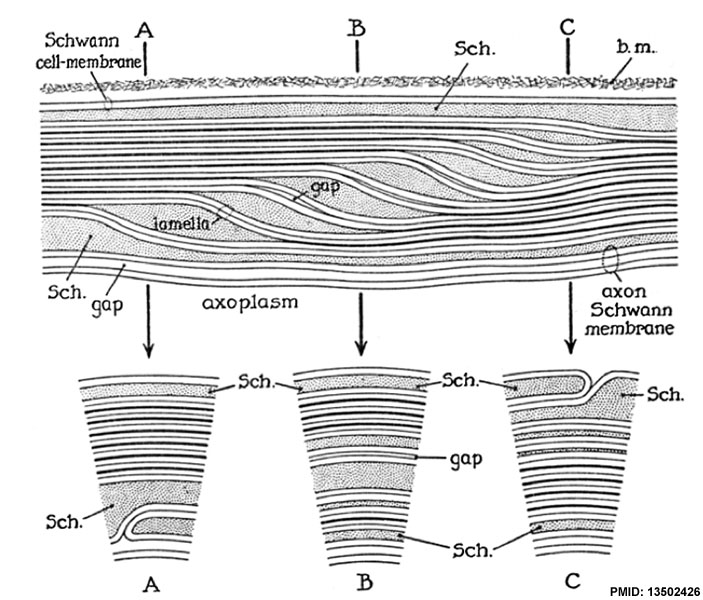
Diagram of a Schmidt-Lanterman cleft in a segment of myelin in longitudinal section
Named after Henry Schmidt and Lanterman (also called Schmidt-Lanterman incisure).
- Schmidt, H. D., Monthly Mitt. J., May 1, 1874,11, 200. (Schmidt, Henry D. 1823-1888 New Orleans pathologist
- Lanterman, A. J., Arch. mikr. Anat., 1877, 13, 1. (American anatomist at Strasbourg)
The outer Schwann cell membrane is shown above as a pair of lines making a unit about 75 A across. Two such units with a gap between them are seen below, making the axon-Schwann membrane. Each myelin lamella is composed of two such units in contact. The heavy dense lines are produced where the adjacent lamellae are in contact with one another. The lamellae each traverse the cleft after separating at the heavy dense lines. Within the clefts the units composing the lamellae sometimes separate slightly with a gap < 100 A wide between them.
This gap is produced by a splitting of the intraperiod line which bisects each myelin lamella (using lamella to mean the structures from center-to-center of each heavy or "major" dense line of compact myelin). The separations between the lamellae in the cleft are much wider than the intralamellar gaps.
At the positions indicated by the letters A, B, and C the appearance of segments of cross-sections of the cleft is shown. In A and C inner and outer mesaxons are shown, respectively. The basement membrane layer (b.m.) is indicated above, but not below.
Reference
ROBERTSON JD. (1958). The ultrastructure of Schmidt-Lanterman clefts and related shearing defects of the myelin sheath. J Biophys Biochem Cytol , 4, 39-46. PMID: 13502426
Copyright
Rockefeller University Press - Copyright Policy This article is distributed under the terms of an Attribution–Noncommercial–Share Alike–No Mirror Sites license for the first six months after the publication date (see http://www.jcb.org/misc/terms.shtml). After six months it is available under a Creative Commons License (Attribution–Noncommercial–Share Alike 4.0 Unported license, as described at https://creativecommons.org/licenses/by-nc-sa/4.0/ ). (More? Help:Copyright Tutorial)
Cite this page: Hill, M.A. (2024, April 23) Embryology Schmidt-Lanterman cleft cartoon.jpg. Retrieved from https://embryology.med.unsw.edu.au/embryology/index.php/File:Schmidt-Lanterman_cleft_cartoon.jpg
- © Dr Mark Hill 2024, UNSW Embryology ISBN: 978 0 7334 2609 4 - UNSW CRICOS Provider Code No. 00098G
File history
Click on a date/time to view the file as it appeared at that time.
| Date/Time | Thumbnail | Dimensions | User | Comment | |
|---|---|---|---|---|---|
| current | 08:56, 15 October 2012 | | 703 × 600 (91 KB) | Z8600021 (talk | contribs) | ==Diagram of a Schmidt-Lanterman cleft in a segment of myelin in longitudinal section== The outer Schwann cell membrane is shown above as a pair of lines making a unit about 75 A across. Two such units with a gap between them are seen below, making the a |
You cannot overwrite this file.
File usage
The following 3 pages use this file: