File:Salvi1898 fig01-15.jpg

Original file (2,500 × 1,836 pixels, file size: 535 KB, MIME type: image/jpeg)
Fig. 1 - 15
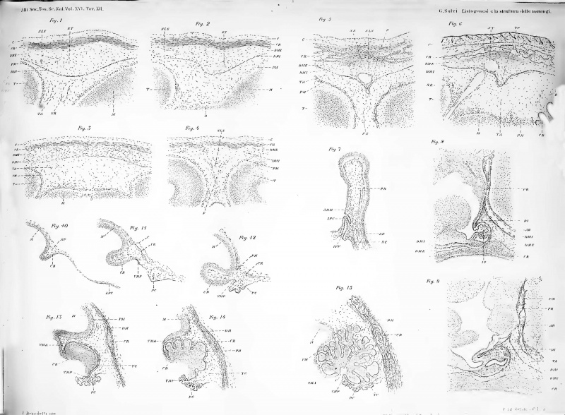
Fig. 1. Median sagittal section of the head of a long guinea pig embryo 20 mm at the groove which remains at the developmental stage between the telencephalon (T) and the midbrain (M).
The sketch of the skull (C) externally limits the primitive meninge and the pia mater (PM) s' is differentiated in the innermost layer. The outline of the dura mater is represented by the DMI straterello and by the layer of tissue interposed between it and the skull (DM E). The arachnoid is represented by the T layer A.
The outer layer of the dura mater includes the veins that will become the superior longitudinal sinus (SLS) and the transverse sinus (ST); in the arachnoid tissue, the right breast (SR).
Fig. 2. Section of a guinea pig embryo of 24 mm. The outer layer of the dura mater begins to become fibrous, and the arachnoid tissue, lax. The hard mother surrounding the transverse breast, protrudes somewhat in the furrow and constitutes the draft of the tentorium.
Fig. 3. Frontal section of the head of a guinea pig embryo of 24 mm through the posterior end of the hemispheres. The sketches of the 3 membranes can be clearly seen.
Fig. 4. Section of a guinea pig embryo of 25 mm towards half of the hemispheres. The inner layer of the dura mater protrudes into the sagittal fissure to constitute the sketch of the scythe of the brain.
Fig. 5. Section of a guinea pig embryo of 58 mm. The scythe followed to develop in the form of a fold of the dural layer, which takes advantage of the arachnoid tissue of the fissure (F).
Fig. 6. Median sagittal section of the head of a guinea pig embryo of 58 mm. The draft of the tentorium (T) develops, in the form of a fold of the dural layer, behind the transverse sinus (ST) and the sinus (SR). The ossification of the skull is already advanced.
Fig. 7. Median sagittal section of the Rathke pillar of a guinea pig embryo of 6 mm. The outline of the pituitary gland protrudes into the embryonic meninges and this form above it is a kind of spur in which it is the diaphragm (ADI).
Fig. 8. Section of the Rathkb pillar region of a guinea pig embryo of 20 mm. The dura mater pushes into the pillar and folds over the pituitary into the diaphragm (DI). The pillar of Eathke is in full involution.
Fig. 9. Section of a guinea pig embryo of 16 mm. The connective tissue interposed between the two laminae of the diaphragm, has also become fibrous, not allowing more to distinguish them.
Fig. 10. Median sagittal section of the head of a guinea pig embryo of mm. 7. The cylindrical epithelium of the choroidal plexus is seen far behind the cerebellar laminae.
Fig. 11. Section of a guinea-pig of 11 mm. The meningeal extension of the choroidal plexuses appears.
Fig. 12. Section of a guinea-pig of 14 mm. The prolongation of the choroidal plexuses is very close to the cerebellum and more developed.
Fig. 13. Section of a guinea-pig of 24 mm. The cerebellum protrudes to the surface of the brain and the sketch of the choroidal canvas (TC) appears.
Fig. 14. Section of a guinea-pig of 36 mm. The facts presented above become more evident and at the same time what becomes the temptation of Kòlliker appears.
Fig. 15. Section of a guinea-pig of 58 mm. The choroidal tissue (TC) is fully developed, and Kolliker's temptation has given rise to the pious mother and the arachnoid of the encephalic regions of the brain.
| Historic Disclaimer - information about historic embryology pages |
|---|
 |
Original Legends
Fig. 1. Sezione sagittale mediana della testa di un embrione di cavia lungo 20 mm. 20 in corrispondenza del solco che a tale stadio di sviluppo rimane fra telencefalo (T) e mesencefalo (M).
L'abbozzo del cranio (C) limita esternamente la meninge primitiva e la pia madre (PM) s' è differenziata nello strato più interno. L'abbozzo della dura madre è rappresentato dallo straterello D M I e dallo strato di tessuto interposto fra questo e il cranio (DM E). L'aracnoide è rappresentata dallo strato T A.
Nello strato esterno della dura madre sono comprese le vene che diventeranno il seno longitudinale superiore (SLS) ed il seno trasverso (S T) ; nel tessuto aracnoideo, il seno retto (S R).
Fig. 2. Sezione e. s. di un embrione di cavia di mm. 24. Lo strato esterno della dura madre comincia a divenire fibroso, ed il tessuto aracnoideo, lasso. La dura madre contornando il seno trasverso, sporge alquanto nel solco e costituisce l'abbozzo del tentorio.
Fig. 3. Sezione frontale della testa di un embrione di cavia di mm. 24 attraverso l'estremità posteriore degli emisferi. Gli abbozzi delle 3 membrane si vedono distintamente.
Fig. 4. Sezione e. s. di un embrione di cavia di mm. 25 verso la metà degli emisferi. Lo strato interno della dura madre sporge nella scissura sagittale a costituire l'abbozzo della falce del cervello.
Fig. 5. Sezione e. s. di un embrione di cavia di mm. 58. La falce seguita a svilupparsi sotto forma di una piega dello strato durale, che si approfonda nel tessuto aracnoideo della scissura (F).
Fig. 6. Sezione sagittale mediana della testa di un embrione di cavia di mm. 58. L'abbozzo del tentorio (T) si sviluppa, sotto forma di piega dello strato durale, al di dietro del seno trasverso (ST) e del seno retto (SR). L'ossificazione del cranio è già avanzata.
Fig. 7. Sezione sagittale mediana del pilastro di Rathke di un embrione di cavia di mm. 6. L'abbozzo dell'ipofisi sporge nella meninge embrionale e questa forma al di sopra di esso una specie di sprone nel quale è l'abozzo del diaframma (A D I).
Fig. 8. Sezione e. s. della regione del pilastro di Rathkb di un embrione di cavia di mm. 20. La dura madre si spinge nel pilastro e si ripiega al di sopra dell' ipofisi formandole iL diaframma (DI). Il pilastro di Eathke è in piena involuzione.
Fig. 9. Sezione e. s. di un embrione di cavia di mm. 36. Il tessuto connettivo interposto fra le due lamine del diaframma, è divenuto anch' esso fibroso non permettendo più di distinguerle.
Fig. 10. Sezione sagittale mediana della testa di un embrione di cavia di mm. 7. Molto indietro della lamina cerebellare si vede 1' epitelio cilindrico ' dell' abbozzo dei plessi coroidei.
Fig. 11. Sezione e. s. di una cavia di mm. 11. Comparisce il prolungamento meningeo dei plessi coroidei.
Fig. 12. Sezione e. s. di una cavia di mm. 14. Il prolungamento dei plessi coroidei si trova molto ravvicinato al cervelletto e più sviluppato.
Fig. 13. Sezione e. s. di una cavia di mm. 24. Il cervelletto sporge alla superficie dell'encefalo e comparisce l'abbozzo della tela coroidea (TC).
Fig. 14. Sezione e. s. di una cavia di mm. 36. I fatti esposti sopra si rendono più evidenti e nel tempo stesso apparisce ciò che diventa il tentorio di Kòlliker.
Fig. 15. Sezione e. s. di una cavia di mm. 58. La tela coroidea (TC) è pienamente sviluppata, ed il tentorio di Kòlliker ha dato origine alla pia madre ed all' aracnoide delle regioni encefaliche respettive.
Reference
Salvi G. Meninges histogenesis and structure (L'istogenesi E La Struttura Delle Meningi). (1898) Thèse de Paris.
Cite this page: Hill, M.A. (2024, April 19) Embryology Salvi1898 fig01-15.jpg. Retrieved from https://embryology.med.unsw.edu.au/embryology/index.php/File:Salvi1898_fig01-15.jpg
- © Dr Mark Hill 2024, UNSW Embryology ISBN: 978 0 7334 2609 4 - UNSW CRICOS Provider Code No. 00098G
File history
Click on a date/time to view the file as it appeared at that time.
| Date/Time | Thumbnail | Dimensions | User | Comment | |
|---|---|---|---|---|---|
| current | 16:00, 24 August 2018 | | 2,500 × 1,836 (535 KB) | Z8600021 (talk | contribs) |
You cannot overwrite this file.
File usage
The following page uses this file: