File:Sabin1909 fig16.jpg
From Embryology
Sabin1909_fig16.jpg (512 × 438 pixels, file size: 120 KB, MIME type: image/jpeg)
Fig. 16.
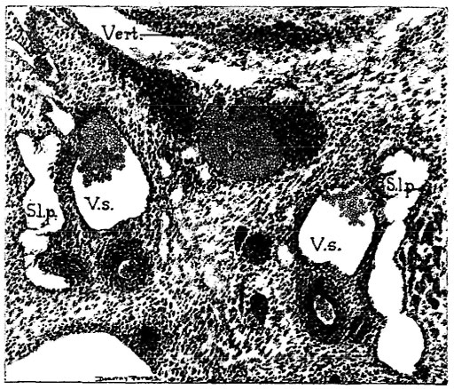
Fig. 16. Coronal section through the posterior lymph sacs as they lie along the primitive sciatic veins, of the same embryo, at a level shown in Fig. 21. X about 49. S. 1. p., saccus lymphaticus posterior; 1’. c., vena candalis; V. s., vena sciatica primitiva.
Reference
Florence R. Sabin, The lymphatic system in human embryos, with a consideration of the morphology of the system as a whole. American Journal of Anatomy Volume 9, Issue 1, pages 43–91, 1909
File history
Click on a date/time to view the file as it appeared at that time.
| Date/Time | Thumbnail | Dimensions | User | Comment | |
|---|---|---|---|---|---|
| current | 15:44, 30 March 2011 |  | 512 × 438 (120 KB) | S8600021 (talk | contribs) | ==Fig. 16.== Fig. 16. Coronal section through the posterior lymph sacs as they lie along the primitive sciatic veins, of the same embryo, at a level shown in Fig. 21. X about 49. S. 1. p., saccus lymphaticus posterior; 1’. c., vena candalis; V. s., ven |
You cannot overwrite this file.
File usage
There are no pages that use this file.
