File:Sabin1909 fig11.jpg

Original file (702 × 1,076 pixels, file size: 109 KB, MIME type: image/jpeg)
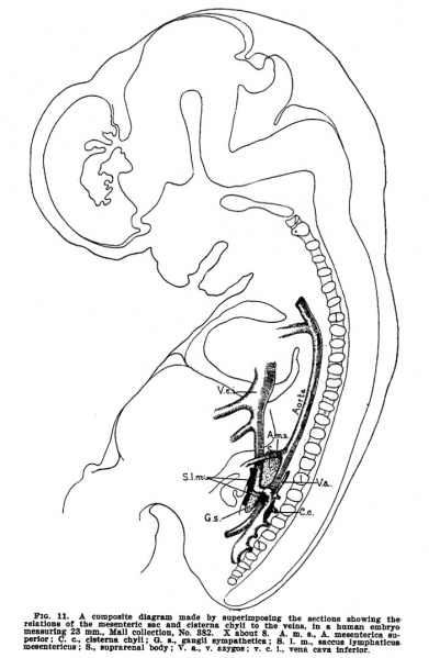
Fig 11. Human embryo measuring 23 mm
Fig 11. A composite diagram made by superimposing the sections showing the relations of the mesenteric sac and cisterna chyli to the veins, in a human embryo measuring 23 mm.
Mall collection, No. 382. X about 3.
cisterna chyli - dilated sac at the lower end of the thoracic duct into which lymph from the intestinal trunk and two lumbar lymphatic trunks flow.
- A. m. s., A. mesenterica superior
- C. c., cisterna chyli
- G. s. gangli sympathetha
- S. l. m.. saccus lymphaticus mesentericus
- S., suprarenal body
- V. a., v. azygos
- v. c. i., vena cava inferior
Approximately Carnegie stage 21 from above drawing and description.
- Links: Lymphatic Development | Sabin
Reference
Sabin FR. The lymphatic system in human embryos, with a consideration of the morphology of the system as a whole. (1909) Amer. J Anat. 9(1): 43–91.
Cite this page: Hill, M.A. (2024, April 24) Embryology Sabin1909 fig11.jpg. Retrieved from https://embryology.med.unsw.edu.au/embryology/index.php/File:Sabin1909_fig11.jpg
- © Dr Mark Hill 2024, UNSW Embryology ISBN: 978 0 7334 2609 4 - UNSW CRICOS Provider Code No. 00098G
File history
Click on a date/time to view the file as it appeared at that time.
| Date/Time | Thumbnail | Dimensions | User | Comment | |
|---|---|---|---|---|---|
| current | 12:50, 30 March 2011 |  | 702 × 1,076 (109 KB) | S8600021 (talk | contribs) | ==Fig 11. Human embryo measuring 23 mm== Fig 11. A composite diagram made by superimposing the sections showing the relations of the mesenteric sac and cisterna chyli to the veins, in a human embryo measuring 23 mm., Mall collection, No. 382. X about 3. |
You cannot overwrite this file.
File usage
The following 5 pages use this file: