File:Respiratorysystem2.png
Respiratorysystem2.png (645 × 575 pixels, file size: 403 KB, MIME type: image/png)
Lower Respiratory Tract
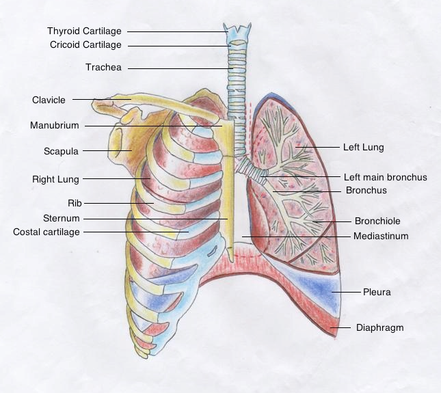
A drawing depicting basic features of the human lower respiratory tract including the thyroid cartilage, rib cage and diaphragm. To reveal the internal structures of the left lung, the skeletal structures and heart have been removed and the left lung has been sectioned in the coronal plane.
Summary
Below are the zones and their contributing structures along with where their originated from.
Conducting zone
|
Respiratory Zone
|
Bones and Muscles
|
Reference
Bibliography Netter, F. (2011). Atlas of human anatomy. Philadelphia, PA: Saunders/Elsevier.
Beginning six months after publication, I, z3333429 grant the public the non-exclusive right to copy, distribute, or display the Work under a Creative Commons Attribution-Noncommercial-Share Alike 3.0 Unported license, as described at http://creativecommons.org/licenses/by-nc-sa/3.0/ and http://creativecommons.org/licenses/by-nc-sa/3.0/legalcode.
--Mark Hill (talk) 10:02, 8 November 2014 (EST) Assessment - this is an excellent student drawing, I have not seen the original so I cannot comment on than aspect. You have also put good information here in the caption. Note that embryonically endoderm does line the entire tract and form the epithelium along its length, so your originating structures are a little simplified. I would also have liked to see a better file name.
- Note - This image was originally uploaded as part of an undergraduate science student project and may contain inaccuracies in either description or acknowledgements. Students have been advised in writing concerning the reuse of content and may accidentally have misunderstood the original terms of use. If image reuse on this non-commercial educational site infringes your existing copyright, please contact the site editor for immediate removal.
File history
Click on a date/time to view the file as it appeared at that time.
| Date/Time | Thumbnail | Dimensions | User | Comment | |
|---|---|---|---|---|---|
| current | 13:49, 24 October 2014 |  | 645 × 575 (403 KB) | Z3333429 (talk | contribs) | ==Lower Respiratory Tract== A drawing depicting basic features of the human lower respiratory tract including the thyroid cartilage, rib cage and diaphragm. To reveal the internal structures of the left lung, the skeletal structures and heart have been... |
You cannot overwrite this file.
File usage
The following 2 pages use this file:
