File:Respiratorysystem1.jpeg
Original file (678 × 960 pixels, file size: 116 KB, MIME type: image/jpeg)
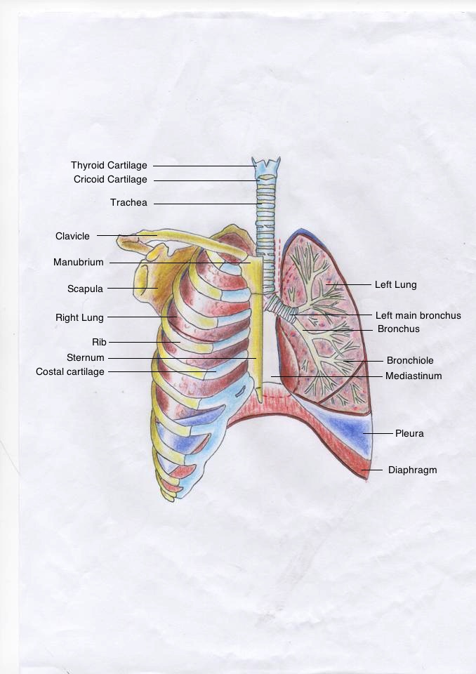
Basic Drawing of Adult Respiratory System
A drawing depicting basic features of the adult human respiratory system from the trachea to the bronchioles and also including the rib cage and diaphragm. To reveal the internal structures of the left lung, the skeletal structures and heart have been removed and the lung has been drawn as a coronal section.
Reference
Bibliography Netter, F. (2011). Atlas of human anatomy. Philadelphia, PA: Saunders/Elsevier.
Beginning six months after publication, I, z3333429 grant the public the non-exclusive right to copy, distribute, or display the Work under a Creative Commons Attribution-Noncommercial-Share Alike 3.0 Unported license, as described at http://creativecommons.org/licenses/by-nc-sa/3.0/ and http://creativecommons.org/licenses/by-nc-sa/3.0/legalcode.
- Note - This image was originally uploaded as part of an undergraduate science student project and may contain inaccuracies in either description or acknowledgements. Students have been advised in writing concerning the reuse of content and may accidentally have misunderstood the original terms of use. If image reuse on this non-commercial educational site infringes your existing copyright, please contact the site editor for immediate removal.
File history
Click on a date/time to view the file as it appeared at that time.
| Date/Time | Thumbnail | Dimensions | User | Comment | |
|---|---|---|---|---|---|
| current | 06:09, 24 October 2014 | | 678 × 960 (116 KB) | Z3333429 (talk | contribs) | ==Basic Drawing of Adult Respiratory System== A drawing depicting basic features of the adult human respiratory system from the trachea to the bronchioles and also including the rib cage and diaphragm. To reveal the internal structures of the left lung... |
You cannot overwrite this file.
File usage
There are no pages that use this file.