File:Renal - S-shaped body stage.jpg

Original file (1,155 × 432 pixels, file size: 99 KB, MIME type: image/jpeg)
Renal Development S-Shaped Body
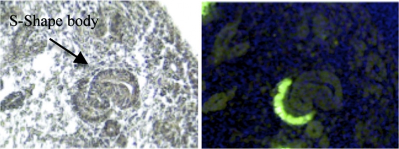
Image shows the S-shaped body stage of renal nephron development in mouse kidney (E15.5)
- Left hand image - S-shaped body.
- Right hand image - MafB-GFP mice show restricted expression of GFP in podocytes. At this stage of development the immature podocytes form a single layer of cells adjacent to the glomerular cleft. As development progresses a capillary loop forms within the cleft and the early glomerulus is encircled by podocytes.
- Links: Renal System Development | Image - Renal - podocyte development | Image - S-shaped body stage | Image - early glomerulus
Figure 1. Journal.pone.0024640.g001.png doi:10.1371/journal.pone.0024640.g001 (images cropped from full figure)
Reference
Brunskill EW, Georgas K, Rumballe B, Little MH, Potter SS (2011) Defining the Molecular Character of the Developing and Adult Kidney Podocyte. PLoS ONE 6(9): e24640. doi:10.1371/journal.pone.0024640
© 2011 Brunskill et al. This is an open-access article distributed under the terms of the Creative Commons Attribution License, which permits unrestricted use, distribution, and reproduction in any medium, provided the original author and source are credited.
File history
Click on a date/time to view the file as it appeared at that time.
| Date/Time | Thumbnail | Dimensions | User | Comment | |
|---|---|---|---|---|---|
| current | 07:03, 17 September 2011 | 1,155 × 432 (99 KB) | S8600021 (talk | contribs) | ==Renal Development S-Shaped Body== Image shows the S-shaped body stage of renal nephron development in mouse kidney (E15.5) * Left hand image - S-shaped body. * Right hand image - MafB-GFP mice show restricted expression of GFP in podocytes. At this st |
You cannot overwrite this file.
File usage
There are no pages that use this file.