File:Reagan1912 plate03.jpg
From Embryology
Size of this preview: 659 × 599 pixels. Other resolution: 1,435 × 1,305 pixels.
Original file (1,435 × 1,305 pixels, file size: 154 KB, MIME type: image/jpeg)
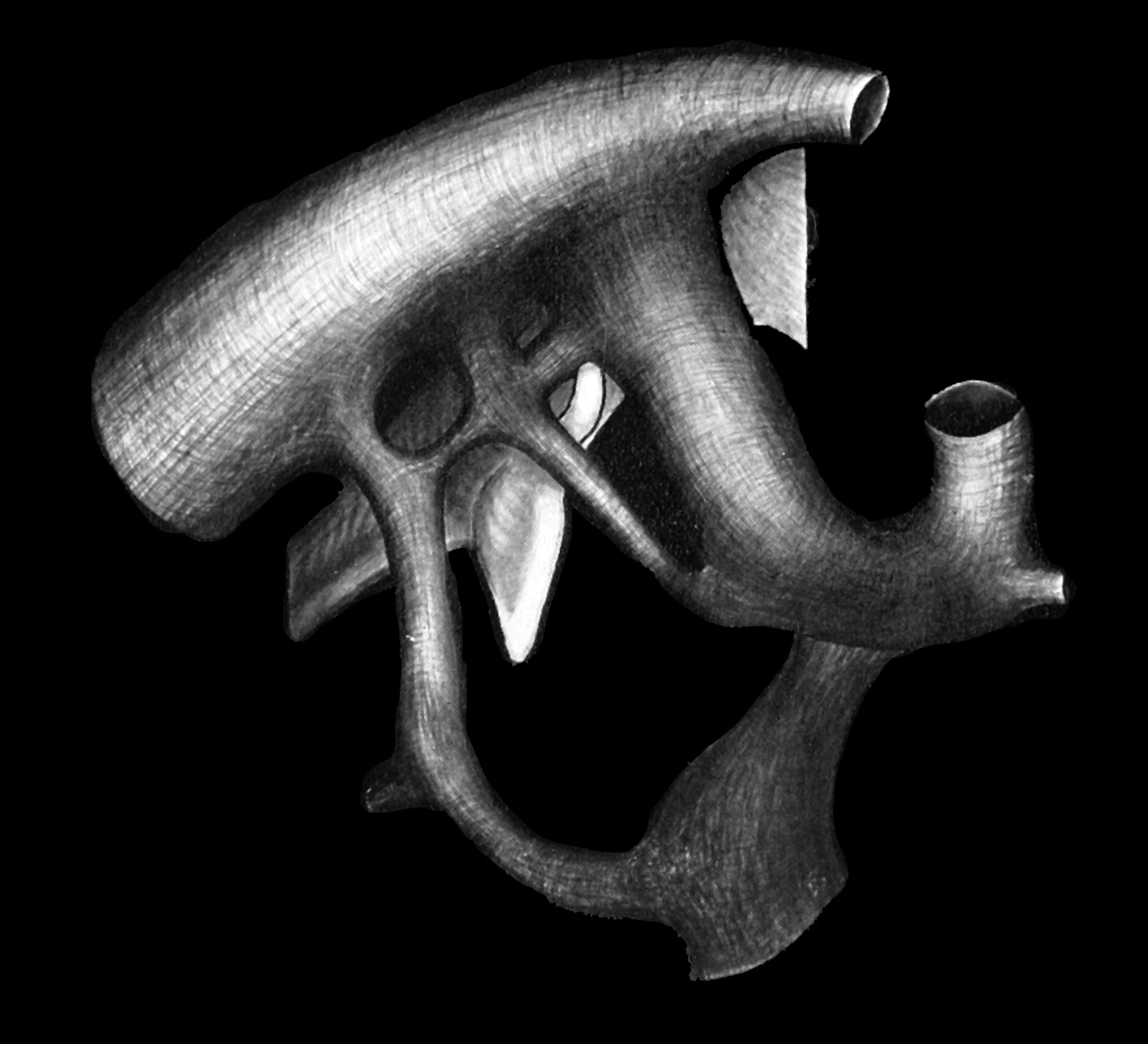
Plate 3
Fig. 8 Reconstruction of the aortic arches of the right side of a 9 mm. pig Series no. 1299, University of Chicago collection.
| Historic Disclaimer - information about historic embryology pages |
|---|
 |
Reference
Reagan F. The fifth aortic arch of mammalian embryos; the nature of the last pharyngeal evagination. (1912) Amer. J Anat. 12(4): 493-505.
Cite this page: Hill, M.A. (2024, April 19) Embryology Reagan1912 plate03.jpg. Retrieved from https://embryology.med.unsw.edu.au/embryology/index.php/File:Reagan1912_plate03.jpg
- © Dr Mark Hill 2024, UNSW Embryology ISBN: 978 0 7334 2609 4 - UNSW CRICOS Provider Code No. 00098G
File history
Click on a date/time to view the file as it appeared at that time.
| Date/Time | Thumbnail | Dimensions | User | Comment | |
|---|---|---|---|---|---|
| current | 13:17, 13 June 2016 | | 1,435 × 1,305 (154 KB) | Z8600021 (talk | contribs) | |
| 13:13, 13 June 2016 | 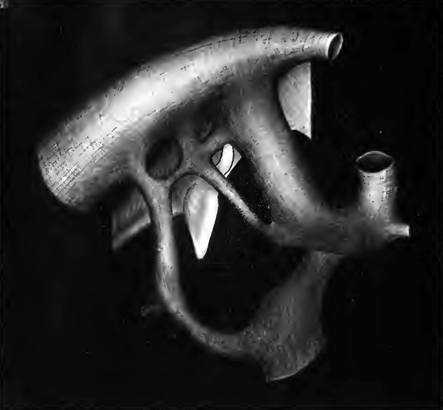 | 1,444 × 1,336 (118 KB) | Z8600021 (talk | contribs) |
You cannot overwrite this file.
File usage
The following page uses this file: