File:Rathke cleft cyst 01.jpg
From Embryology
Rathke_cleft_cyst_01.jpg (493 × 600 pixels, file size: 46 KB, MIME type: image/jpeg)
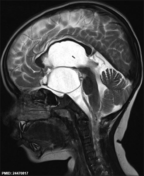
Rathke's cleft Cyst MRI
Magnetic resonance imaging (MRI) sagittal section is showing suprasellar Rathke's cleft cyst (RCC) with normal compressed pituitary.
Reference
<pubmed>24470817</pubmed>| J Pediatr Neurosci.
Copyright
http://creativecommons.org/licenses/by-nc-sa/3.0/
Figure 2
File history
Click on a date/time to view the file as it appeared at that time.
| Date/Time | Thumbnail | Dimensions | User | Comment | |
|---|---|---|---|---|---|
| current | 15:19, 3 September 2014 |  | 493 × 600 (46 KB) | Z8600021 (talk | contribs) | ==Rathke's cleft Cyst MRI== Magnetic resonance imaging (MRI) sagittal section is showing suprasellar Rathke's cleft cyst (RCC) with normal compressed pituitary. ===Reference=== <pubmed>24470817</pubmed>| [http://www.pediatricneurosciences.com/articl... |
You cannot overwrite this file.
File usage
There are no pages that use this file.
