File:Ramsey1960-fig01.jpg
Original file (1,000 × 638 pixels, file size: 175 KB, MIME type: image/jpeg)
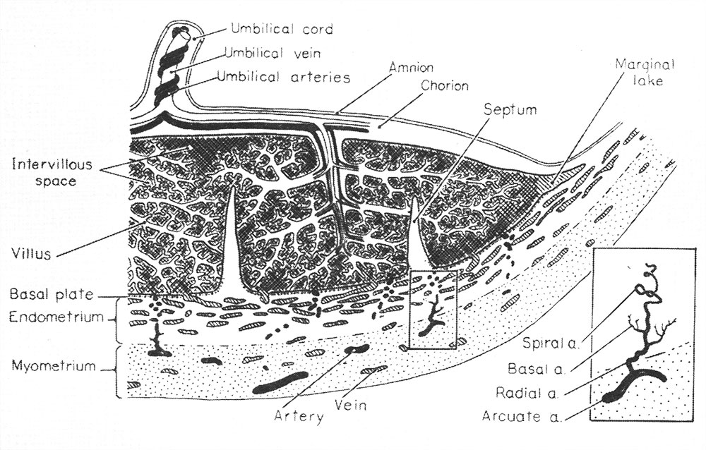
Fig. 1. Diagrammatic representation of a portion of the primate (hemochorial) placenta attached to the uterine wall
The inset shows, at higher magnification, the components of a single arterial stem.
(From Ramsey,8 in Report of 15th M&R Paediatric Research Conference 1955.)
Reference
Ramsey EM. Corner GW. Jr. Donner MW. and Stran HM. Radioangiographic studies of circulation in the maternal placenta of the rhesus monkey: preliminary report. (1960) Proc. Natl. Acad. Sci. U.S.A., 46(7): 1003-8 PMID 16590693
Copyright
Proceedings National Academy of Sciences (PNAS) Liberalization of PNAS copyright policy: Noncommercial use freely allowed Note original Author should be contacted for permission to reuse for Educational purposes. See also PNAS Author Rights and Permission FAQs
- Cozzarelli NR, Fulton KR, Sullenberger DM. Liberalization of PNAS copyright policy: noncommercial use freely allowed. Proc Natl Acad Sci U S A. 2004 Aug 24;101(34):12399. PMID15314225 "Our guiding principle is that, while PNAS retains copyright, anyone can make noncommercial use of work in PNAS without asking our permission, provided that the original source is cited."
Cite this page: Hill, M.A. (2024, April 25) Embryology Ramsey1960-fig01.jpg. Retrieved from https://embryology.med.unsw.edu.au/embryology/index.php/File:Ramsey1960-fig01.jpg
- © Dr Mark Hill 2024, UNSW Embryology ISBN: 978 0 7334 2609 4 - UNSW CRICOS Provider Code No. 00098G
File history
Click on a date/time to view the file as it appeared at that time.
| Date/Time | Thumbnail | Dimensions | User | Comment | |
|---|---|---|---|---|---|
| current | 09:40, 13 May 2016 | | 1,000 × 638 (175 KB) | Z8600021 (talk | contribs) | |
| 09:40, 13 May 2016 | 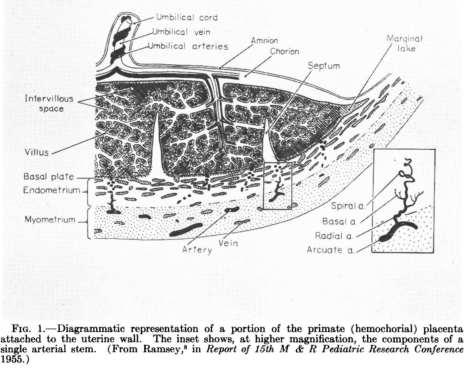 | 1,509 × 1,195 (446 KB) | Z8600021 (talk | contribs) | ==Fig. 1. Diagrammatic representation of a portion of the primate (hemochorial) placenta attached to the uterine wall== The inset shows, at higher magnification, the components of a single arterial stem. (From Ramsey,8 in Report of 15th M&R Paediatr... |
You cannot overwrite this file.
File usage
The following 3 pages use this file: