File:Radford1908 fig05.jpg
Radford1908_fig05.jpg (789 × 524 pixels, file size: 48 KB, MIME type: image/jpeg)
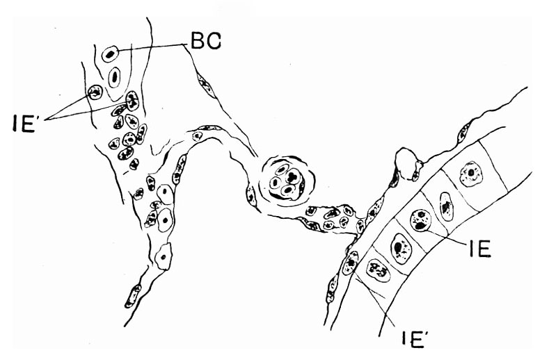
Fig. 5. From an embryo of 15 mm total length
Portion of intestinal wall, with small terminal branch of mesenteric artery and mesentery. White and red blood-corpuscles, mesenchyme cells in the mesentery, and strayed endodermal cells. One cell from the intestinal epithelium lies between it and the epithelium of the mesentery.
BC, blood-corpuscles ; IE, intestinal epithelium; IE’, strayed endodermal cells.
Reference
Radford M. Development of the spleen. (1908) J Anat Physiol. 42: 288-301.
Cite this page: Hill, M.A. (2024, April 23) Embryology Radford1908 fig05.jpg. Retrieved from https://embryology.med.unsw.edu.au/embryology/index.php/File:Radford1908_fig05.jpg
- © Dr Mark Hill 2024, UNSW Embryology ISBN: 978 0 7334 2609 4 - UNSW CRICOS Provider Code No. 00098G
File history
Click on a date/time to view the file as it appeared at that time.
| Date/Time | Thumbnail | Dimensions | User | Comment | |
|---|---|---|---|---|---|
| current | 13:04, 19 July 2019 | | 789 × 524 (48 KB) | Z8600021 (talk | contribs) | |
| 13:02, 19 July 2019 |  | 1,118 × 847 (128 KB) | Z8600021 (talk | contribs) |
You cannot overwrite this file.
File usage
The following page uses this file: