File:Promising System for Selecting Healthy In Vitro Fertilized Embryos in Cattle.png

Original file (2,067 × 1,238 pixels, file size: 1 MB, MIME type: image/png)
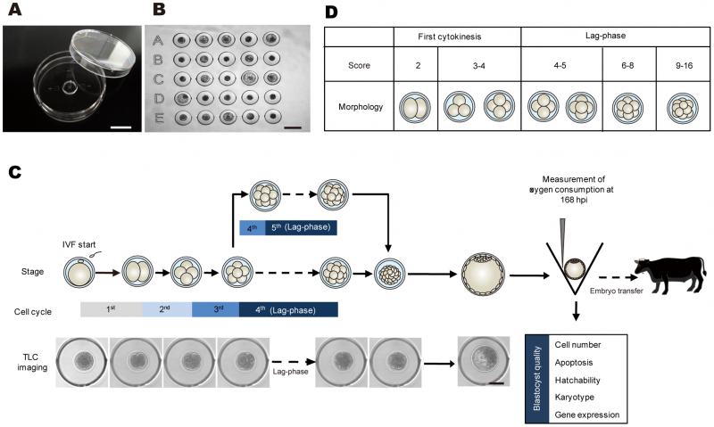
Overview of the experimental scheme for selecting healthy in vitro fertilized embryos in cattle
In vitro development of bovine IVF embryos in the developed microwell culture dish with identification code (A and B) was tracked with time-lapse cinematography (TLC) for 168 h post-insemination (hpi). SECM was used to measure oxygen consumption in embryos that developed to the blastocyst stage at 168 hpi (C). Timing of the of the first cleavage; duration of the second, third, and lag-phases resulting in developmental arrest at the fourth or fifth cell cycle; number of blastomeres; presence or absence of multiple fragments; unevenness or evenness of division at the end of the first cleavage and at the onset of the lag-phase; cell cycle observed lag-phase (fourth or fifth cell cycle); and oxygen consumption at 168 hpi were examined in relation to blastocyst qualities such as cell number (n = 173), apoptosis incidence (n = 74), hatchability (n = 195), karyotype (n = 111), and gene expression (n = 75) with multivariate analysis. Based on the identified prognostic factors that reflected the various blastocyst qualities, OPU-IVF blastocysts (n = 52) were then assessed and transferred to recipient cows (n = 52). (D) The number of blastomeres at the end of the first cleavage was categorized as 2 and 3/4 blastomeres. The numbers of blastomeres at the onset of the lag-phase were categorized as 4/5, 6–8, and 9–16 based on the result from Fig. S1. Bar = 10 mm (A), 300 µm (B) and 100 µm (C).
--Mark Hill (talk) 16:07, 21 August 2014 (EST) I have made some minor changes to the summary information formatting. Please use this format in future.
Reference
<pubmed>22590579</pubmed>
Copyright
© 2012 Sugimura et al. This is an open-access article distributed under the terms of the Creative Commons Attribution License, which permits unrestricted use, distribution, and reproduction in any medium, provided the original author and source are credited.
- Note - This image was originally uploaded as part of an undergraduate science student project and may contain inaccuracies in either description or acknowledgements. Students have been advised in writing concerning the reuse of content and may accidentally have misunderstood the original terms of use. If image reuse on this non-commercial educational site infringes your existing copyright, please contact the site editor for immediate removal.
File history
Click on a date/time to view the file as it appeared at that time.
| Date/Time | Thumbnail | Dimensions | User | Comment | |
|---|---|---|---|---|---|
| current | 10:33, 20 August 2014 | | 2,067 × 1,238 (1 MB) | Z3418989 (talk | contribs) | In vitro development of bovine IVF embryos in the developed microwell culture dish with identification code (A and B) was tracked with time-lapse cinematography (TLC) for 168 h post-insemination (hpi). SECM was used to measure oxygen consumption in emb... |
You cannot overwrite this file.
File usage
The following page uses this file: