File:Prentiss1906 fig04.jpg
Original file (1,618 × 1,064 pixels, file size: 490 KB, MIME type: image/jpeg)
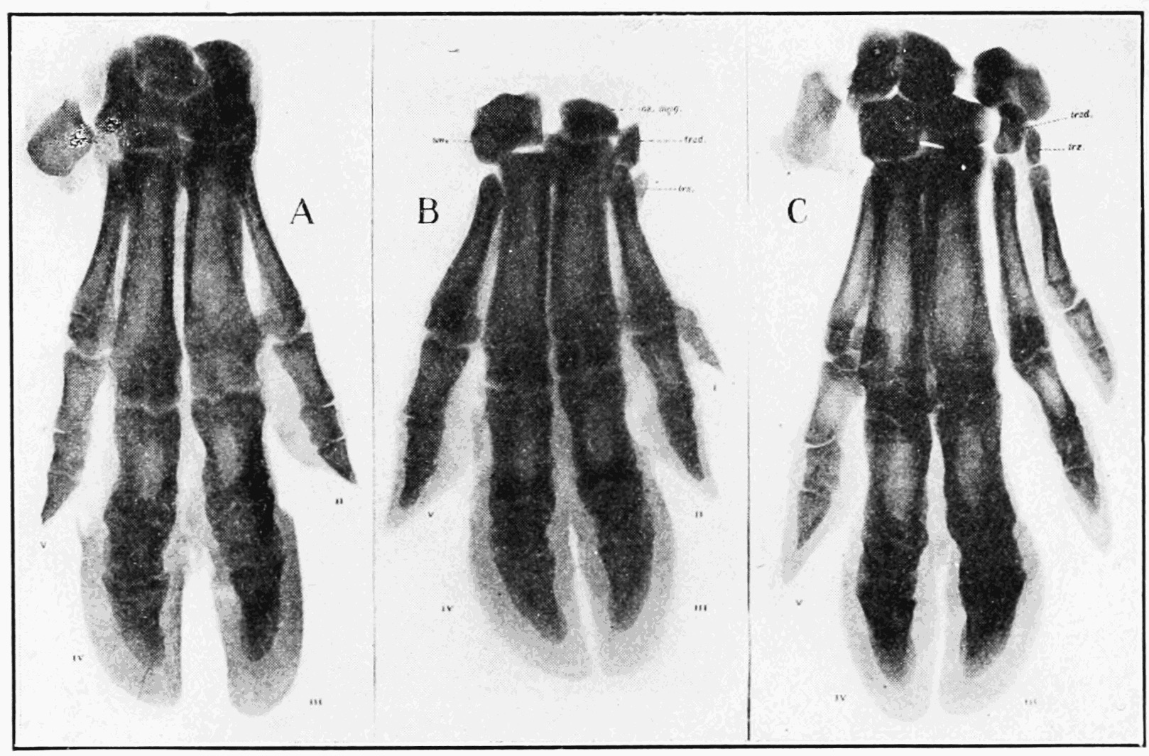
Fig. 4. X-ray photographs of the pig's manus
Showing normal structure and reversionary polydactylism.
A, bones of normal manus;
B, manus in which the pollex is represented by two phalanges and the distal end of the metacarpal bone (I.);
C, manus with pollex completely developed; trz., trapezium.
fig 1 | fig 2 | fig 3 | fig 4 | fig 5 | fig 6
| fig 7
| Historic Disclaimer - information about historic embryology pages |
|---|
 |
Reference
Prentiss CW. Extra digits and digital reductions. (1906) Popular Science Monthly. 336-448.
Cite this page: Hill, M.A. (2024, April 25) Embryology Prentiss1906 fig04.jpg. Retrieved from https://embryology.med.unsw.edu.au/embryology/index.php/File:Prentiss1906_fig04.jpg
- © Dr Mark Hill 2024, UNSW Embryology ISBN: 978 0 7334 2609 4 - UNSW CRICOS Provider Code No. 00098G
File history
Click on a date/time to view the file as it appeared at that time.
| Date/Time | Thumbnail | Dimensions | User | Comment | |
|---|---|---|---|---|---|
| current | 09:36, 9 April 2019 | | 1,618 × 1,064 (490 KB) | Z8600021 (talk | contribs) |
You cannot overwrite this file.
File usage
The following page uses this file: