File:Prentiss001.jpg
From Embryology
Prentiss001.jpg (599 × 567 pixels, file size: 86 KB, MIME type: image/jpeg)
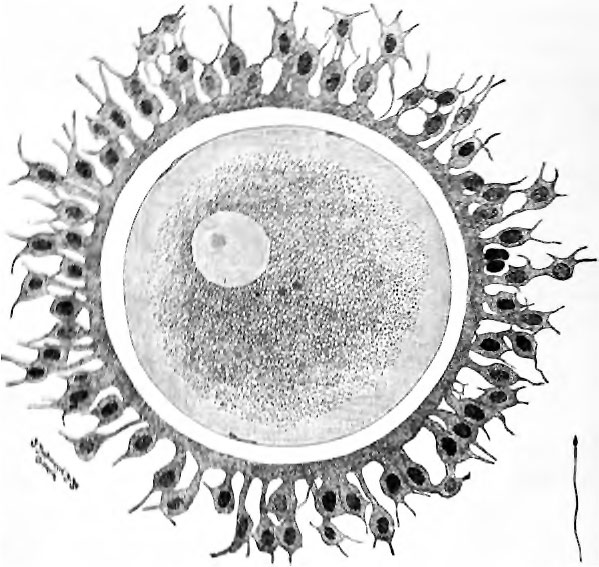
Fig. 1. Human ovum examined fresh in the liquor folliculi
(Waldeyer) original x415
The zona pellucida is seen as a thick clear girdle surrounded by the cells of the corona radiate. The nearly mature egg itself shows a central granular cytoplasmic area and a peripheral clear layer, and encloses the nucleus in which is seen the nucleolus. At the right is a spermatozoon correspondingly enlarged.
| Historic Disclaimer - information about historic embryology pages |
|---|
| Pages where the terms "Historic" (textbooks, papers, people, recommendations) appear on this site, and sections within pages where this disclaimer appears, indicate that the content and scientific understanding are specific to the time of publication. This means that while some scientific descriptions are still accurate, the terminology and interpretation of the developmental mechanisms reflect the understanding at the time of original publication and those of the preceding periods, these terms, interpretations and recommendations may not reflect our current scientific understanding. (More? Embryology History | Historic Embryology Papers) |
Alaboratorymanu01prengoog_0023-fig-1.jpg
File history
Click on a date/time to view the file as it appeared at that time.
| Date/Time | Thumbnail | Dimensions | User | Comment | |
|---|---|---|---|---|---|
| current | 08:18, 25 October 2011 |  | 599 × 567 (86 KB) | S8600021 (talk | contribs) | Prentiss Alaboratorymanu01prengoog_0023-fig-1.jpg |
You cannot overwrite this file.
File usage
The following page uses this file:

