File:Placental imaging 03A.jpg
Original file (800 × 738 pixels, file size: 181 KB, MIME type: image/jpeg)
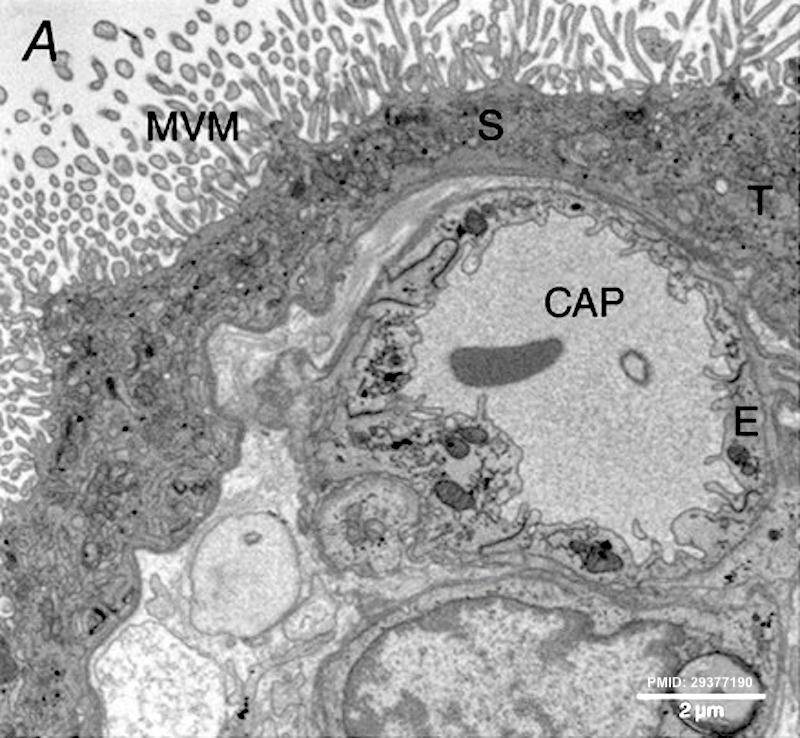
Placental Imaging
A transmission electron micrograph of terminal villi showing
MVM - microvillous membrane
CAP - underlying capillary
T - trophoblast
E - endothelium
- Links: placenta | trophoblast | Fig 3 | Haemomonochorial human placenta (EM)
Reference
Nye GA, Ingram E, Johnstone ED, Jensen OE, Schneider H, Lewis RM, Chernyavsky IL & Brownbill P. (2018). Human placental oxygenation in late gestation: experimental and theoretical approaches. J. Physiol. (Lond.) , 596, 5523-5534. PMID: 29377190 DOI.
Copyright
© 2018 University of Oxford. The Journal of Physiology published by John Wiley & Sons Ltd on behalf of The Physiological Society
This is an open access article under the terms of the Creative Commons Attribution License, which permits use, distribution and reproduction in any medium, provided the original work is properly cited.
Figure 3. Evaluating imaging techniques for use in assessing placental structure Tjp12830-fig-0003-m.jpg
Panel A cropped from Fig 3, resized, contrast adjusted and PMID label added.
Cite this page: Hill, M.A. (2024, April 24) Embryology Placental imaging 03A.jpg. Retrieved from https://embryology.med.unsw.edu.au/embryology/index.php/File:Placental_imaging_03A.jpg
- © Dr Mark Hill 2024, UNSW Embryology ISBN: 978 0 7334 2609 4 - UNSW CRICOS Provider Code No. 00098G
File history
Click on a date/time to view the file as it appeared at that time.
| Date/Time | Thumbnail | Dimensions | User | Comment | |
|---|---|---|---|---|---|
| current | 11:18, 10 April 2019 | | 800 × 738 (181 KB) | Z8600021 (talk | contribs) | ==Placental Imaging== A transmission electron micrograph of terminal villi showing microvillous membrane (MVM), an underlying capillary (CAP), a syncytiotrophoblast (S), a trophoblast (T) and endothelium (E). :'''Links:''' {{placenta}} | [[:File:Pl... |
You cannot overwrite this file.
File usage
The following page uses this file: