File:Placental chorioangioma ultrasound 01.jpg
Original file (1,183 × 837 pixels, file size: 101 KB, MIME type: image/jpeg)
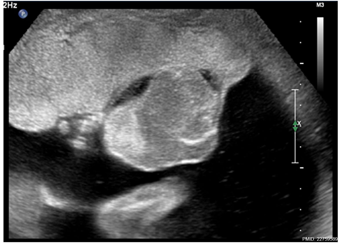
Placental Chorioangioma Ultrasound
An ultrasound examination revealed normal fetal growth for gestational age. Her amniotic fluid index was 48 cm, with the deepest pocket of 13 cm and no signs of fetal hydrops. Ultrasound middle cerebral artery peak systolic velocity (MCA PSV) color Doppler was 48 cm/s, which was 1.71 Multiple of Median (MoM) for gestational age. The placenta was implanted anteriorly with a detectable vascularized tumor measuring 42 mm × 56 mm × 58 mm with a noticeable feeding vessel at the root.
Gross examination (Gross image 1) of the placenta revealed a yellowish, well-circumscribed firm mass measuring 5 cm × 5 cm connected by two vessels to the placenta.
Histopathologic examination revealed a placental disc 15 cm × 17 cm × 13 cm, with a three-vessel umbilical cord that was attached peripherally and measured 9 cm × 1.5 cm. The weight of the placenta was 530 g. The tumor was confirmed to be a chorioangioma.
- Links: Ultrasound scan | Ultrasound blood flow | Gross image 1 | Gross image 2 | Gross image 3 | Placental Chorioangioma | Placenta - Abnormalities
Reference
<pubmed>22759589</pubmed>| PMC3419096 | J Med Case Rep
Copyright
© 2012 Babic et al.; licensee BioMed Central Ltd. This is an Open Access article distributed under the terms of the Creative Commons Attribution License (http://creativecommons.org/licenses/by/2.0), which permits unrestricted use, distribution, and reproduction in any medium, provided the original work is properly cited.
Figure 5. Original image was adjusted in size, contrast and labelling.
1752-1947-6-183-1.jpg
File history
Click on a date/time to view the file as it appeared at that time.
| Date/Time | Thumbnail | Dimensions | User | Comment | |
|---|---|---|---|---|---|
| current | 11:19, 7 June 2014 | | 1,183 × 837 (101 KB) | Z8600021 (talk | contribs) | 1752-1947-6-183-1.jpg |
You cannot overwrite this file.
File usage
The following page uses this file: