File:Placenta cord arteries abnormality 02.jpg
Placenta_cord_arteries_abnormality_02.jpg (752 × 332 pixels, file size: 47 KB, MIME type: image/jpeg)
Placenta cord arteries abnormality
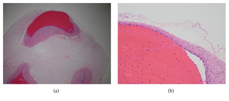
(a). Low power photomicrograph of section of umbilical cord showing superficial location of the umbilical artery (top) with absence of overlying Wharton’s jelly, attenuated vascular media, and degeneration of the amnion. This is in contrast to the umbilical vein (lower left) and other umbilical artery (lower right) both of which were completely surrounded by Wharton’s jelly and amnion (complete views not in the field). H &E, X 20. (b). High power photomicrograph of ulcerated umbilical artery demonstrating markedly thinned media, absent Wharton’s jelly, and degeneration of the overlying amnion. H &E, X 200.
Reference
Curtin WM, Maines JL, DeAngelis CT, Condon NA, Ural SH & Millington KA. (2019). Hemorrhage from Umbilical Cord Ulceration Identified on Real-Time Ultrasound in a Fetus with Duodenal Atresia. Case Rep Obstet Gynecol , 2019, 2680170. PMID: 30906606 DOI.
Copyright
© 2019 William M. Curtin et al. This is an open access article distributed under the Creative Commons Attribution License, which permits unrestricted use, distribution, and reproduction in any medium, provided the original work is properly cited.
Figure 3. CRIOG2019-2680170.003.jpg
File history
Click on a date/time to view the file as it appeared at that time.
| Date/Time | Thumbnail | Dimensions | User | Comment | |
|---|---|---|---|---|---|
| current | 15:58, 16 April 2019 |  | 752 × 332 (47 KB) | Z8600021 (talk | contribs) |
You cannot overwrite this file.
File usage
There are no pages that use this file.
