File:Pig lung alveolarization.jpg
From Embryology
Pig_lung_alveolarization.jpg (600 × 389 pixels, file size: 38 KB, MIME type: image/jpeg)
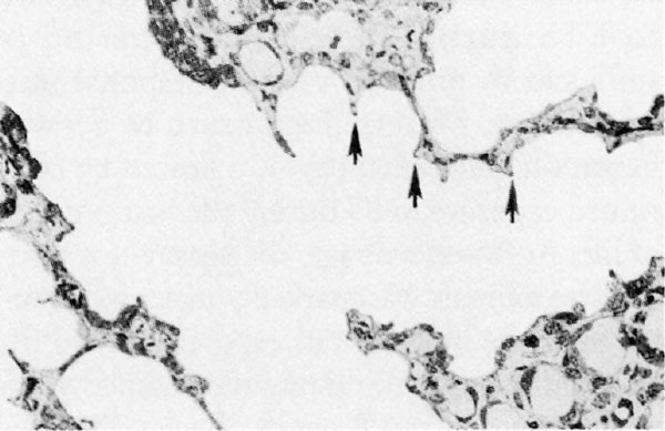
Fetal Pig Lung Alveolarization
Centriacinar region from the fetal lung of a near-term pig with the parenchymal walls undergoing the process of alveolarization.
Short buds of secondary alveolar septa (arows) extend out from thick primary alveolar septa to form a future alveolar duct.
In all species, alveolarization continues postnatally.
Reference
<pubmed>10852845</pubmed>| PMC1637815 | Environ Health Perspect.
Environmental Health Perspectives Vol 108, Supplement 3 June 2000
File history
Click on a date/time to view the file as it appeared at that time.
| Date/Time | Thumbnail | Dimensions | User | Comment | |
|---|---|---|---|---|---|
| current | 09:44, 1 March 2011 |  | 600 × 389 (38 KB) | S8600021 (talk | contribs) | ==Fetal Pig Lung Alveolarization== Centriacinar region from the fetal lung of a near-term pig with the parenchymal walls undergoing the process of alveolarization. Short buds of secondary alveolar septa (arows) extend out from thick primary alveolar s |
You cannot overwrite this file.
File usage
There are no pages that use this file.
