File:Pharyngeal arch segmentation model - Tbx and Fox.jpg

Original file (1,509 × 783 pixels, file size: 163 KB, MIME type: image/jpeg)
Summary
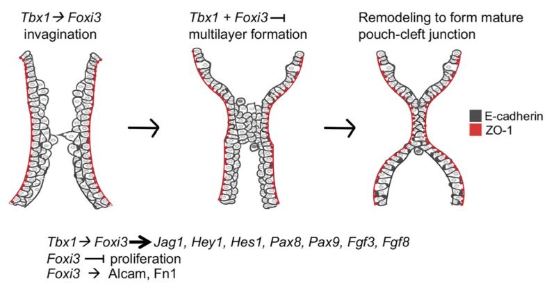
Tbx1 and Foxi3 functions in PA segmentation
Cartoon of epithelial cells during PA segmentation for each arch. Tbx1 and Foxi3 are co-expressed where segmentation to individual pharyngeal arches occurs. When segmentation of the distal PA begins at E8.5, invagination of E-cadherin expressing cells (dark gray) is initiated and a partially stratified multilayer of epithelium forms by E8.75-E9.0, near the point where invagination continues. Tbx1 acts upstream of Foxi3 to promote proper invagination as indicated. The outermost cell layer maintains apical/ basal polarity as depicted by ZO-1 expression (red), while the inner layers do not. The epithelial cells begin to project towards each other as shown. Next, a multilayer is juxtaposed where invagination has advanced, and a zippering process ensues from the center of the region, where cells are reorganized. Finally, a dual-layer of intercalated epithelial cells are formed at the mature pouch-cleft junction and both arches are separated. Foxi3 appears to inhibit excess proliferation of PE cells early, while Tbx1 doesn’t alter proliferation. In Tbx1 null mutant embryos more cells are present in the shortened PA. Inactivation of Tbx1 or Foxi3 results in the appearance of excessive layers of endoderm cells, in particular, where invagination is initiating. Therefore, these genes might both promote invagination and restrict excessive multilayers during PA segmentation. We found that Tbx1 and Foxi3 may act in the same pathway upstream of some Notch pathway genes, Pax8 and Pax9, as well as Fgf3. Some of these genes might be required pharyngeal segmentation. It is previously known that Tbx1 and Foxi3 act upstream of Fgf8. While loss of Foxi3 resulted in reduction of Alcam and Fibronectin expression in the extracellular matrix, global loss of Tbx1 did not have the same role. Thus, this data explains, in part, the basis of the genetic interaction between the two genes.
Reference
Hasten E & Morrow BE. (2019). Tbx1 and Foxi3 genetically interact in the pharyngeal pouch endoderm in a mouse model for 22q11.2 deletion syndrome. PLoS Genet. , 15, e1008301. PMID: 31412026 DOI.
Copyright
© 2019 Hasten, Morrow. This is an open access article distributed under the terms of the Creative Commons Attribution License, which permits unrestricted use, distribution, and reproduction in any medium, provided the original author and source are credited.
Cite this page: Hill, M.A. (2024, April 25) Embryology Pharyngeal arch segmentation model - Tbx and Fox.jpg. Retrieved from https://embryology.med.unsw.edu.au/embryology/index.php/File:Pharyngeal_arch_segmentation_model_-_Tbx_and_Fox.jpg
- © Dr Mark Hill 2024, UNSW Embryology ISBN: 978 0 7334 2609 4 - UNSW CRICOS Provider Code No. 00098G
File history
Click on a date/time to view the file as it appeared at that time.
| Date/Time | Thumbnail | Dimensions | User | Comment | |
|---|---|---|---|---|---|
| current | 01:33, 10 March 2020 |  | 1,509 × 783 (163 KB) | Z8600021 (talk | contribs) | ==Tbx1 and Foxi3 functions in PA segmentation== Cartoon of epithelial cells during PA segmentation for each arch. Tbx1 and Foxi3 are co-expressed where segmentation to individual pharyngeal arches occurs. When segmentation of the distal PA begins at E8.5, invagination of E-cadherin expressing cells (dark gray) is initiated and a partially stratified multilayer of epithelium forms by E8.75-E9.0, near the point where invagination continues. Tbx1 acts upstream of Foxi3 to promote proper invagin... |
You cannot overwrite this file.
File usage
The following 3 pages use this file: