File:Peyers patches ileocolonoscopy 01.jpg
Peyers_patches_ileocolonoscopy_01.jpg (600 × 482 pixels, file size: 45 KB, MIME type: image/jpeg)
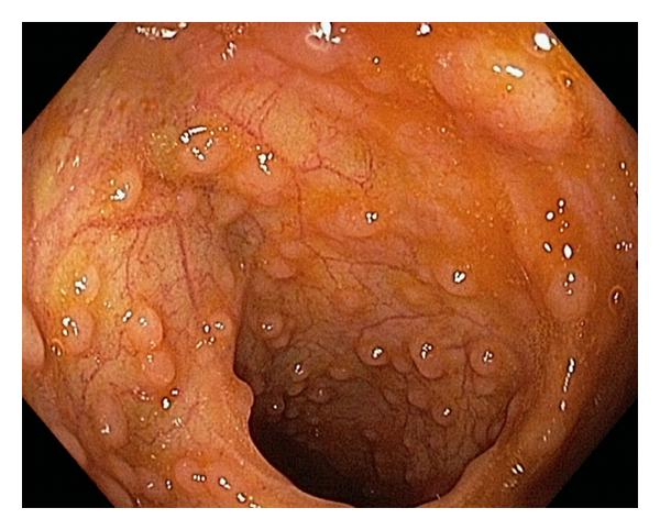
Peyer’s patches in the Distal Ileum
Peyer’s patches form a lymphoid ring in the distal ileum.
Seen in a 20-years-old man during ileocolonoscopy.
Reference
Jung C, Hugot JP & Barreau F. (2010). Peyer's Patches: The Immune Sensors of the Intestine. Int J Inflam , 2010, 823710. PMID: 21188221 DOI.
Copyright
© 2010 Camille Jung et al. This is an open access article distributed under the Creative Commons Attribution License, which permits unrestricted use, distribution, and reproduction in any medium, provided the original work is properly cited.
823710.fig.001.jpg
Cite this page: Hill, M.A. (2024, April 24) Embryology Peyers patches ileocolonoscopy 01.jpg. Retrieved from https://embryology.med.unsw.edu.au/embryology/index.php/File:Peyers_patches_ileocolonoscopy_01.jpg
- © Dr Mark Hill 2024, UNSW Embryology ISBN: 978 0 7334 2609 4 - UNSW CRICOS Provider Code No. 00098G
File history
Click on a date/time to view the file as it appeared at that time.
| Date/Time | Thumbnail | Dimensions | User | Comment | |
|---|---|---|---|---|---|
| current | 15:30, 25 February 2015 |  | 600 × 482 (45 KB) | Z8600021 (talk | contribs) | 823710.fig.001.jpg |
You cannot overwrite this file.
File usage
The following page uses this file:
