File:Patten1938 text-fig04.jpg

Original file (1,000 × 1,432 pixels, file size: 316 KB, MIME type: image/jpeg)
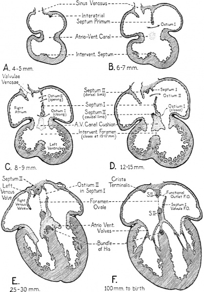
Text-Fig. 4. Sectional plans of the embryonic heart in the frontal plane
Showing extent of growth of the various cardiac septa at several stages of development. These diagrams give specifically for the human embryo a more precise picture of the rate of progress of partitioning than do the schematic drawings of Text-Figs. 1-3.
Stippled areas in the diagrams indicate the distribution of endocardial connective tissue, muscle is shown in diagonal hatching, and the epicardium in solid black. The lightly stippled areas in the atrioventricular canal in B and C indicate the lomtion of the dorsal and ventral endocardial cushions of the atrioventricular canal before they have grown suficiently to fuse with each other in the plane of the diagram.
Reference
Patten BM. Developmental defects at the foramen ovale. (1938) Am J Pathol. 14(2):135-162. PMID 19970381
Cite this page: Hill, M.A. (2024, April 25) Embryology Patten1938 text-fig04.jpg. Retrieved from https://embryology.med.unsw.edu.au/embryology/index.php/File:Patten1938_text-fig04.jpg
- © Dr Mark Hill 2024, UNSW Embryology ISBN: 978 0 7334 2609 4 - UNSW CRICOS Provider Code No. 00098G
File history
Click on a date/time to view the file as it appeared at that time.
| Date/Time | Thumbnail | Dimensions | User | Comment | |
|---|---|---|---|---|---|
| current | 16:01, 27 February 2017 | | 1,000 × 1,432 (316 KB) | Z8600021 (talk | contribs) | |
| 16:00, 27 February 2017 |  | 1,310 × 2,326 (503 KB) | Z8600021 (talk | contribs) |
You cannot overwrite this file.
File usage
The following page uses this file: