File:Patten1938 plate32.jpg
Original file (1,280 × 1,928 pixels, file size: 321 KB, MIME type: image/jpeg)
Plate 32
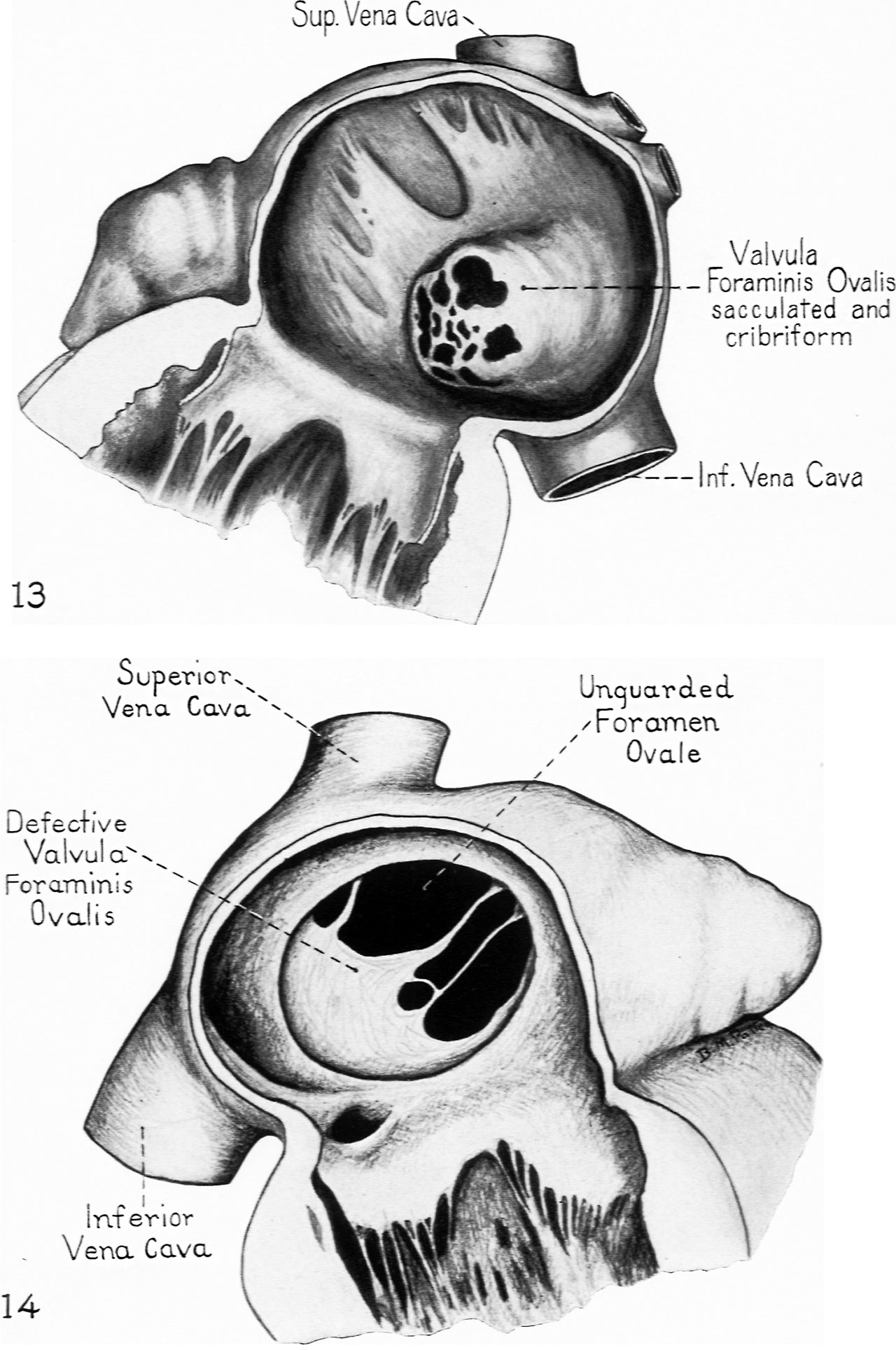
FIG. 13. Valvula foraminis ovalis markedly sacculated toward left atriinn and showing multiple perforations of considerable size. No clinical history. Dissecting room specimen “from an old man” sent in by Dr. John Donaldson. University of Pittsburgh.
FIG. 14. Drawn from specimen No. 3027, Pathologisch-Anatomisches Institut. Vienna. The heart was from a charwoman who died suddenly of pulmonary thrombosis at the age of 52 years. Rokitansky (I 75, p. 52) gives a brief unillustrated record of the case. The heart was “very large, 90 mm. long and 115 mm. broad” with rormded apex. The similarity of the morphological picture presented by this adult heart and the infant heart shown in Fig. II is interesting.
Reference
Patten BM. Developmental defects at the foramen ovale. (1938) Am J Pathol. 14(2):135-162. PMID 19970381
Cite this page: Hill, M.A. (2024, April 23) Embryology Patten1938 plate32.jpg. Retrieved from https://embryology.med.unsw.edu.au/embryology/index.php/File:Patten1938_plate32.jpg
- © Dr Mark Hill 2024, UNSW Embryology ISBN: 978 0 7334 2609 4 - UNSW CRICOS Provider Code No. 00098G
File history
Click on a date/time to view the file as it appeared at that time.
| Date/Time | Thumbnail | Dimensions | User | Comment | |
|---|---|---|---|---|---|
| current | 16:15, 27 February 2017 | | 1,280 × 1,928 (321 KB) | Z8600021 (talk | contribs) | |
| 16:14, 27 February 2017 |  | 1,538 × 2,478 (413 KB) | Z8600021 (talk | contribs) |
You cannot overwrite this file.
File usage
The following 2 pages use this file: