File:Patten1938 plate31.jpg
Original file (1,280 × 1,799 pixels, file size: 236 KB, MIME type: image/jpeg)
Plate 31
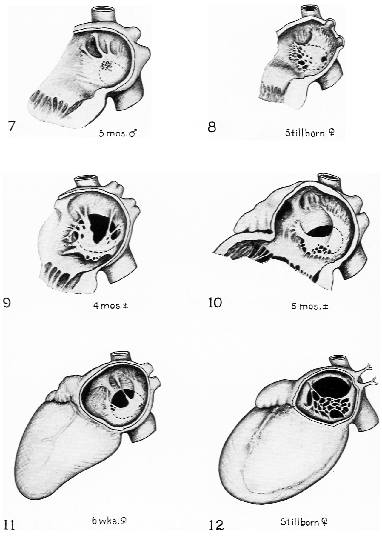
FIG. 7. Multiple small perforations of valvula. The formation of ostium secundum normally starts with the appearance of small openings which later coalesce. Here such openings have appeared in a definitely abnormal location.
FIG. 8. Case similar to that shovm in Fig. 7. except that the openings are larger and more vddely distributed.
FIGS. 9. 10 and 11. Various combinations of marginal over resorption of the types shown in Figs. 1-6 with small multiple perforations similar to those shown in Figs. 7 and 8.
FIG. 12. Extreme resorption of valvula combined with abnormally large foramen ovale due to defective development of septum secundum. There is also in this heart unbalanced development of the ventricles. the left ventricle being very small, correlated probably with a defective puhnonary circuit as indicated by the marked stenosis of the pulmonary veins.
Reference
Patten BM. Developmental defects at the foramen ovale. (1938) Am J Pathol. 14(2):135-162. PMID 19970381
Cite this page: Hill, M.A. (2024, April 19) Embryology Patten1938 plate31.jpg. Retrieved from https://embryology.med.unsw.edu.au/embryology/index.php/File:Patten1938_plate31.jpg
- © Dr Mark Hill 2024, UNSW Embryology ISBN: 978 0 7334 2609 4 - UNSW CRICOS Provider Code No. 00098G
File history
Click on a date/time to view the file as it appeared at that time.
| Date/Time | Thumbnail | Dimensions | User | Comment | |
|---|---|---|---|---|---|
| current | 16:11, 27 February 2017 | | 1,280 × 1,799 (236 KB) | Z8600021 (talk | contribs) | |
| 16:10, 27 February 2017 | 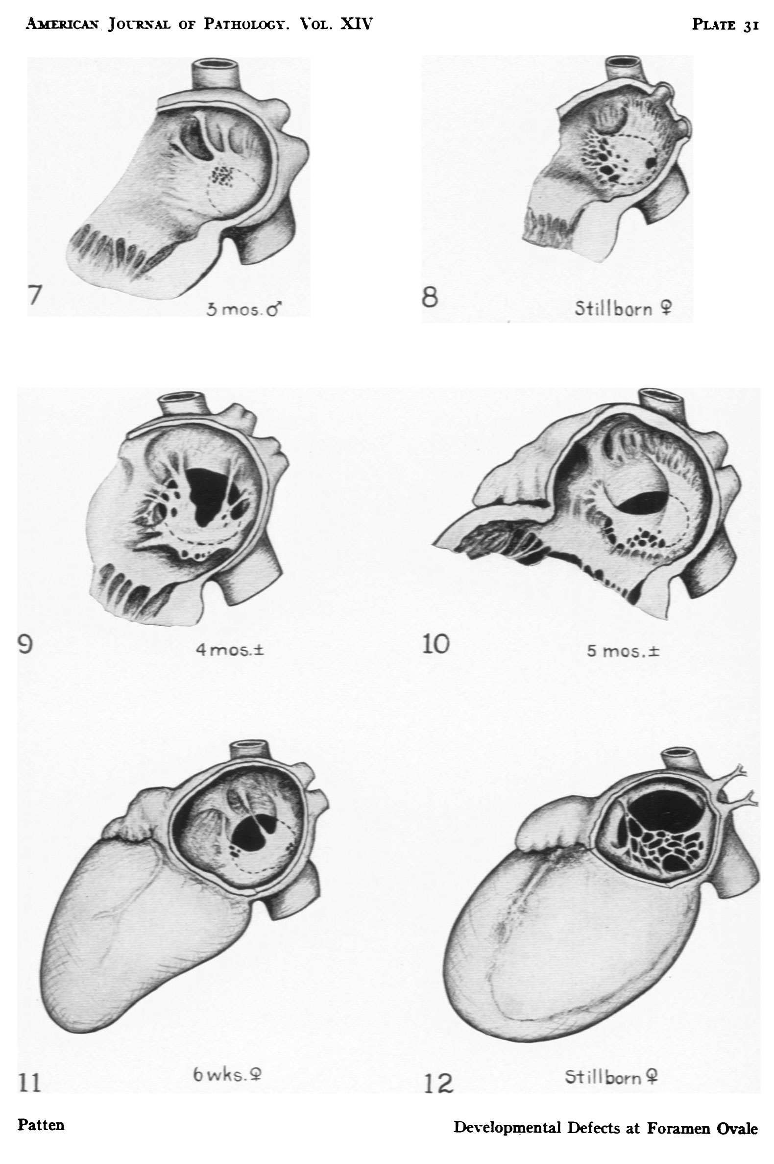 | 1,580 × 2,374 (325 KB) | Z8600021 (talk | contribs) |
You cannot overwrite this file.
File usage
The following 2 pages use this file: