File:Patten054.jpg
Original file (764 × 1,060 pixels, file size: 179 KB, MIME type: image/jpeg)
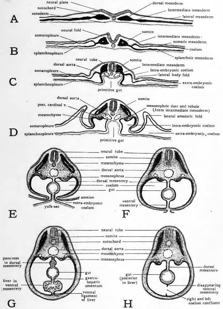
Fig. 54. Schematic diagrams of cross sections at various stages to show the establishment of the coelom and mesenteries
The portion of the coelom which gives rise to the embryonic body cavities is first marked off by the series of folds which separate the body of the embryo from the yolk (Fig. 54, C, D). As the closure of the ventral body wall progresses (Fig. 54, E, F) the embryonic coelom becomes completely separated from the extra-embryonic. The delayed closure of the ventral body wall in the yolk-stalk region, results in the embryonic and extra-embryonic coelom retaining their open communication at this point for a long time after they have been completely separated elsewhere.
The same folding process which establishes the vejitral body wall completes the gut ventrally (Fig. 54, C to F) . Meanwhile the right and left ccelomic chambers are expanded mesiad. As a result the newly closed gut comes to lie suspended between the two layers of splanchnic mesoderm which constitute the mesial walls of the right and left coelomic chambers, respectively. The double layers of splanchnic mesoderm which thus become apposed to the gut and support it in the body cavity are known as mesenteries. The mesentery dorsal to the gut, suspending it from the dorsal body wall is the primary dorsal mesentery, and that ventral to the gut, attaching it to the ventral body wall is the primary ventral mesentery.
When the dorsal and ventral mesenteries are first established they constitute a complete membranous partition dividing the body cavity into right and left halves. The primary dorsal mesentery persists in large part but the ventral mesentery early disappears bringing the right and left ccelomic chambers into confluence ventral to the gut and establishing the unpaired condition of the body cavity characteristic of the adult.
In considering the early development of the heart (Chapter IX) the formation of the dorsal and ventral mesocardia was taken up. In their relation to the other mesenteries of the body, the mesocardia are to be regarded as special regions of the primary ventral mesentery. In the most cephalic part of the body cavity, the gut lies embedded in the dorsal body wall instead of being suspended by the primary dorsal mesentery as it is farther caudally (Cf. Fig. 26, E and Fig. 54, F). The ventral mesentery is, however, developed in the same manner anteriorly as it is posteriorly and when the heart is formed it is suspended in the most anterior part of the primary ventral mesentery. The dorsal and ventral mesocardia are the parts of the primary ventral mesentery lying dorsal to the heart, and vontral to the heart, respectively (Fig. 26, D).
When the ventral mesocardium, and a little later the dorsal mesocardium, breaks through, the primary right and left coelomic chambers become confluent to form the pericardial region of the body cavity (Figs. 24 and 55). Later in development the ventral mesentery farther caudally disappears so that caudally as well as cephahcally an unpaired condition of the coelom is brought about (Fig. 54, H).
In the liver region the ventral mesentery does not disappear. The liver arises as an outgrowth from the gut and in its development extends into the ventral mesentery (Fig. 54, G). The portion of the ventral mesentery dorsal to the liver persists as the gastro-hepatic omentum, and the portion ventral to the liver persists as its ventral ligament (falciform ligament) (Fig- 55)
- Links: Introduction | Gametes and Fertilization | Segmentation | Entoderm | Primitive Streak and Mesoderm | Primitive Streak to Somites | 24 Hours | 24 to 33 Hours | 33 to 39 Hours | 40 to 50 Hours | Extra-embryonic Membranes | 50 to 55 Hours | Day 3 to 4 | References | All Figures
| Historic Disclaimer - information about historic embryology pages |
|---|
 |
Reference
Patten BM. The Early Embryology of the Chick. (1920) Philadelphia: P. Blakiston's Son and Co.
Cite this page: Hill, M.A. (2024, April 19) Embryology Patten054.jpg. Retrieved from https://embryology.med.unsw.edu.au/embryology/index.php/File:Patten054.jpg
- © Dr Mark Hill 2024, UNSW Embryology ISBN: 978 0 7334 2609 4 - UNSW CRICOS Provider Code No. 00098G
File history
Click on a date/time to view the file as it appeared at that time.
| Date/Time | Thumbnail | Dimensions | User | Comment | |
|---|---|---|---|---|---|
| current | 02:06, 17 January 2011 | | 764 × 1,060 (179 KB) | S8600021 (talk | contribs) | {{Template:Patten_1920_Figures}} |
You cannot overwrite this file.
File usage
The following 3 pages use this file: