File:Patten049.jpg
Original file (807 × 864 pixels, file size: 119 KB, MIME type: image/jpeg)
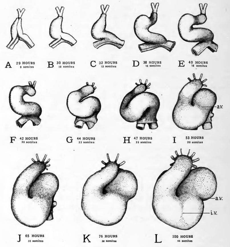
Fig. 49. Ventral views of the heart at various stages to show its changes of shape and its regional differentiation
All the drawings were made from dissections with the aid of camera lucida outlines.
The outer of the two layers shown is the epi-myocardium; the inner, the endocardium. In the stages represented in Figs. E-H torsion of the embryo's body is going on at the level of the heart. Since torsion involves the more cephalic regions first and progresses caudad the transverse axis of the body of the embryo is at different inclinations to the yolk at the cephalic end and at the caudal end of the heart. In drawing these figures their orientation was taken from the body at the level of the conus region of the heart, the sinus region therefore appears inclined.
Abbreviations:
- a.v. - constriction between atrium and ventricle
- i.v. - interventricular groove.
- Links: Introduction | Gametes and Fertilization | Segmentation | Entoderm | Primitive Streak and Mesoderm | Primitive Streak to Somites | 24 Hours | 24 to 33 Hours | 33 to 39 Hours | 40 to 50 Hours | Extra-embryonic Membranes | 50 to 55 Hours | Day 3 to 4 | References | All Figures
| Historic Disclaimer - information about historic embryology pages |
|---|
 |
Reference
Patten BM. The Early Embryology of the Chick. (1920) Philadelphia: P. Blakiston's Son and Co.
Cite this page: Hill, M.A. (2024, April 20) Embryology Patten049.jpg. Retrieved from https://embryology.med.unsw.edu.au/embryology/index.php/File:Patten049.jpg
- © Dr Mark Hill 2024, UNSW Embryology ISBN: 978 0 7334 2609 4 - UNSW CRICOS Provider Code No. 00098G
File history
Click on a date/time to view the file as it appeared at that time.
| Date/Time | Thumbnail | Dimensions | User | Comment | |
|---|---|---|---|---|---|
| current | 02:05, 17 January 2011 | | 807 × 864 (119 KB) | S8600021 (talk | contribs) | {{Template:Patten_1920_Figures}} |
You cannot overwrite this file.
File usage
The following 3 pages use this file: