File:Patten048.jpg
Original file (797 × 857 pixels, file size: 174 KB, MIME type: image/jpeg)
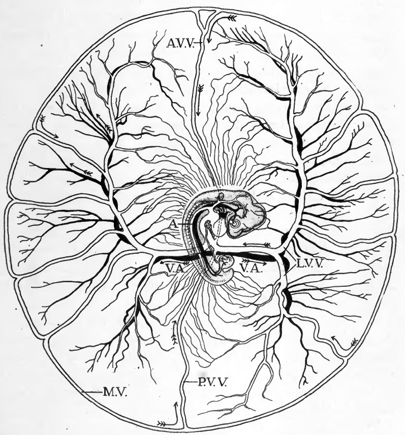
Fig. 48. Diagram to show course of vitelline circulation in chick of about four days
(After Lillie.)
For the intra-embryonic vessels see Fig. 47. The direction of blood flow is indicated by arrows.
Abbreviations:
- A, dorsal aorta
- A.V.V., anterior vitelline vein
- L.V.V., lateral vitelline vein
- M.V., marginal vein (sinus terminalis)
- P.V.V., posterior vitelline vein
- V.A., vitelline artery.
The course of the vitelline circulation in chicks of four days is shown diagrammatically in Figures 47 and 48. Circulating in the rich plexus of small vessels on the yolk, the blood finally makes its way either directly into one or another of the larger vitelline veins, or to the sinus terminalis which acts as a collecting channel, and then over the sinus terminalis to one of the vitelline veins. The vitelline veins conVerge toward the yolk-stalk where they empty into the omphalomesenteric veins. The omphalomesenteric veins at first paired throughout their entire length have been brought together proximally by the closure of the ventral body wall and become fused to form a median vessel within the body of the embryo. It is through this vessel that the vitelline blood eventually reaches the heart. In the heart the blood of the vitelline, intra-embryonic, and allantoic circulations is mingled. The mixed blood passes out by the ventral aorta and the aortic arches into the dorsal aorta. Leaving the dorsal aorta through the vitelline arteries the blood is returned to the yolk-sac.
- Links: Introduction | Gametes and Fertilization | Segmentation | Entoderm | Primitive Streak and Mesoderm | Primitive Streak to Somites | 24 Hours | 24 to 33 Hours | 33 to 39 Hours | 40 to 50 Hours | Extra-embryonic Membranes | 50 to 55 Hours | Day 3 to 4 | References | All Figures
| Historic Disclaimer - information about historic embryology pages |
|---|
 |
Reference
Patten BM. The Early Embryology of the Chick. (1920) Philadelphia: P. Blakiston's Son and Co.
Cite this page: Hill, M.A. (2024, April 25) Embryology Patten048.jpg. Retrieved from https://embryology.med.unsw.edu.au/embryology/index.php/File:Patten048.jpg
- © Dr Mark Hill 2024, UNSW Embryology ISBN: 978 0 7334 2609 4 - UNSW CRICOS Provider Code No. 00098G
File history
Click on a date/time to view the file as it appeared at that time.
| Date/Time | Thumbnail | Dimensions | User | Comment | |
|---|---|---|---|---|---|
| current | 02:04, 17 January 2011 | | 797 × 857 (174 KB) | S8600021 (talk | contribs) | {{Template:Patten_1920_Figures}} |
You cannot overwrite this file.
File usage
The following 3 pages use this file: